Rehabilitation of Trauma Using Dental Implants

The goal of replacing missing teeth while respecting otherwise untouched tooth structure and the avoidance of crown reduction in bridge preparation make the use of dental implants an ideal option for restoring dentitions ravaged by traumatic tooth loss. Patients who suffer tooth loss resulting from traumatic injuries of the dentoalveolar complex can be divided into growing and nongrowing patients. The placement of dental implants can have deleterious effects on the growing alveolar process. The authors’ philosophy is to respect growth and delay implant reconstruction until the cessation of skeletal or alveolar growth, as documented by serial lateral cephalometrics radiographs taken 6 months apart.

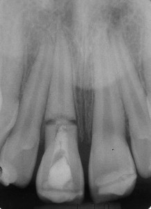
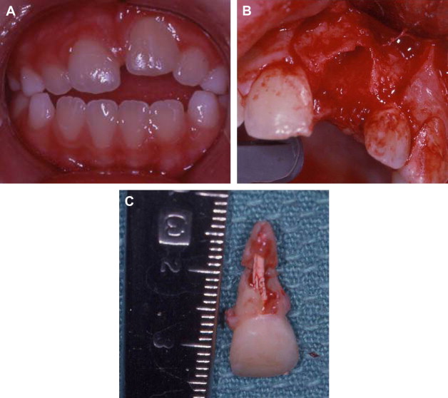
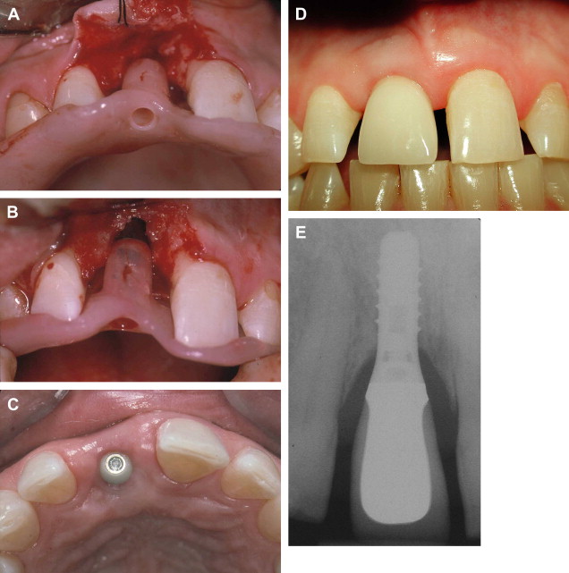
The ability to replace teeth without damaging the residual dentition makes the use of dental implants an ideal option to consider for restoring dentitions ravaged by traumatic tooth loss ( Fig. 1 ). Patients who suffer tooth loss resulting from traumatic injuries of the dentoalveolar complex may still be growing, and, in fact, certain injuries resulting in tooth loss are seen more commonly in young patients (for example, avulsions of anterior teeth). Dental implants, like ankylosed teeth, can have deleterious effects on the growing alveolar process ( Fig. 2 A–C). As a general rule, the authors’ philosophy is to respect growth by delaying implant placement until the cessation of skeletal growth ( Fig. 3 A–E), as documented by serial lateral cephalometric radiographs taken 6 months apart.

Fig. 1
Transverse root fracture of traumatized maxillary central incisor with poor prognosis. Treatment planning of this case must take into consideration the current state of growth and damage to surrounding teeth and their prognoses.

Fig. 2
( A ) Ankylosed maxillary permanent central incisor that had been avulsed, replanted, and treated with root canal therapy. The ankylosed tooth has inhibited alveolar growth and has become submerged below the occlusal plane of the surrounding dentition. ( B ) Alveolar defect noted at the time of removal of the ankylosed permanent maxillary central incisor undergoing inflammatory root resorption. ( C ) Specimen of ankylosed permanent maxillary central incisor with evidence of inflammatory root resorption. This patient was still growing so that dental implant placement was delayed until growth cessation had been attained.

Fig. 3
( A ) Permanent maxillary right central incisor lost in an ice hockey–related accident in a nongrowing patient. A prefabricated stent is used to communicate to the surgeon the vertical, buccolingual and mesiodistal positioning and axial inclination desired by the prosthodontist. ( B ) The stent is used to check the correct vertical positioning of the implant fixture margin. ( C ) Correctly positioned dental implant seen from the occlusal aspect after 3 months of healing. ( D ) Restored implant after 8 years of follow-up. ( E ) Periapical radiograph of restored dental implant after 8 years of follow-up.

In addition to growth, a number of other factors associated with tooth loss also must be considered when assessing a traumatized dentition ( Box 1 ).

Box 1

  • Cessation of skeletal growth

  • Single versus multiple tooth loss

  • Prognosis of residual dentition and likelihood of future tooth loss

  • Associated loss of alveolar bone

  • Associated soft tissue scarring and deficits

  • Associated fractures of the facial bones

Factors to consider with implant-assisted reconstruction of the traumatized dental alveolus

Assessment of the residual dentition

No two complex fractures involving the dentition, it seems, are ever the same. The posttraumatic residual dentition must be analyzed carefully to help predict its prognosis. Teeth that have been reimplanted, suffered root fractures, or been treated endodontically will impact adversely the overall prognosis (see Fig. 2 A–C), as they may be lost in the future, necessitating further costly treatment. Considerations of potential future tooth loss will often have important medicolegal implications especially as it related to insurance liability.

Loss of hard and soft tissues

Loss of alveolar bone associated with tooth avulsion may require that ridge augmentation, perhaps using guided bone regeneration or other techniques, be incorporated into the reconstruction treatment plan ( Fig. 4 A–C). In addition, the deleterious effects of soft tissue scarring on the final esthetic outcome must be anticipated and communicated to the patient and family ( Fig. 5 A–C) and dealt with where appropriate through the use of soft tissue grafting or flaps ( Fig. 6 A–H).

Fig. 4
( A ) Traumatic loss of permanent maxillary left lateral incisor, canine, and first and second premolars 6 months after a boating accident. The buccolingual dimension of the residual alveolar ridge appears undiminished and the soft tissues seem to be well healed. ( B ) Panoramic radiograph shows dental implant placement. ( C ) Note elongated prosthetic crowns on the restored left side of the maxilla, compared with the natural crowns on the right, necessitated by vertically deficient alveolar bone.

Fig. 5
( A ) Nongrowing individual who suffered dentoalveolar trauma in a motor vehicle accident 9 months before with loss of permanent maxillary left central, lateral, canine, and first premolar teeth. The buccolingual ridge width seems adequate on the occlusal view. ( B ) Three implant fixtures have been used to replace the missing teeth. ( C ) Frontal view of the crowns supported by the three dental implant fixtures 9 months after their insertion. Note the residual posttraumatic scarring of the gingival mucosa. Also note the blunted papillae between the implant-supported crowns. Another option would have been to place only two implant fixtures and span the edentulous segment with a fixed bridge, which we now recognize may have resulted in a more ideal formation of the papillae.

Fig. 6
( A ) Connective tissue graft harvested from palatal mucosa to restore soft tissue bulk associated with permanent teeth at the left maxillary lateral incisor and canine sites. ( B ) The harvested connective tissue graft. ( C ) The connective tissue graft is inserted into a pocket created on the labial aspect of the implants at the lateral incisor and canine sites. ( D ) The healed site 3 months after graft placement seen from the labial aspect. ( E ) The healed site seen from the occlusal view. ( F ) Model showing the position of the healed implant fixtures. ( G ) Occlusal view of the restored implants. ( H ) Labial view of the restored implants and the surrounding soft tissues.

Loss of hard and soft tissues

Loss of alveolar bone associated with tooth avulsion may require that ridge augmentation, perhaps using guided bone regeneration or other techniques, be incorporated into the reconstruction treatment plan ( Fig. 4 A–C). In addition, the deleterious effects of soft tissue scarring on the final esthetic outcome must be anticipated and communicated to the patient and family ( Fig. 5 A–C) and dealt with where appropriate through the use of soft tissue grafting or flaps ( Fig. 6 A–H).

Jan 23, 2017 | Posted by in Oral and Maxillofacial Surgery | Comments Off on Rehabilitation of Trauma Using Dental Implants

VIDEdental - Online dental courses

Get VIDEdental app for watching clinical videos