During childhood, the anterior maxilla is susceptible to injury, and the loss of incisors is one of the most serious injuries. In many cases, autotransplantation is the best alternative for children who lose an incisor during the growth phase. This case report describes the treatment of a boy who had a traumatic injury when he was 8 years old that resulted in avulsion of the maxillary right central incisor. When he sought treatment at age 10, the space was lost as was bone in the incisor region. Because he lacked space in the mandibular arch for proper tooth alignment, extractions were planned. One extracted premolar was transplanted into the space of the missing maxillary incisor area. The posttreatment results were good, and follow-up records 7 and 9 years after treatment showed healthy periodontal support and cortical bone gain in the transplanted tooth’s buccal area.
Highlights
- •
A 10-year-old boy, physically healthy, with history of dental trauma and absence of the maxillary right central incisor, was evaluated.
- •
The selected treatment was to transplant the mandibular right second premolar to the maxillary right central incisor region, where there was bone loss.
- •
Results showed cortical bone gain on the transplanted tooth’s buccal root, and 9-year follow-up radiography showed healthy periodontal support tissue.
The incisor root development phase coincides with the period of childhood in which trauma occurs frequently in the anterior maxilla. During this age, tooth avulsion occurs because the incisor roots are still growing and do not have good bone implantation. However, there are situations where the trauma is of such a magnitude that reimplantation is not possible. Consequently, treatment becomes limited because it is contraindicated to place dental implants or other restorative treatment while the patient still has the potential for alveolar growth.
The transplantation of immature premolars has become a viable method for restoring edentulous areas because they can maintain the function and preserve the alveolar crest; this is advantageous for young patients when compared with dental implants that are static and do not erupt to compensate for future growth.
The success of autotransplanted teeth is associated with periodontal ligament healing, no root resorption, bone and gum healing, and pulp and root development. To maintain this sequence of factors, atraumatic surgery is fundamental, and multidisciplinary participation is necessary to allow for a functional and esthetic restoration.
Recent advances in computerized tomography have allowed for the visualization of structures that were impossible to evaluate in the past. Through these new technologies, the visualization of the periodontal ligament and new bone formation has become a reality.
This case report presents the autotransplantation of a premolar to the region of a central incisor through surgery with preservation of the periodontal ligament. The receiver site exhibited considerable bone resorption because tooth loss occurred 2 years before the initiation of treatment. The preservation of dental structures (cementum and periodontal ligament) allowed for bone formation around the transplanted tooth. This finding was possible only with the use of 3-dimensional tomography as a means of observation.
Diagnosis and etiology
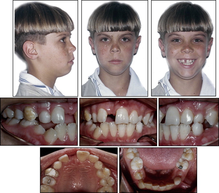
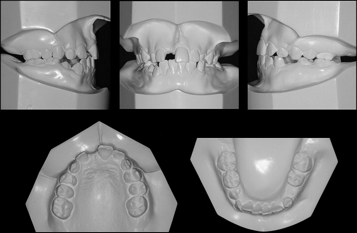
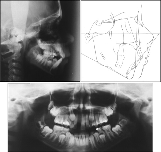
A 10-year-old boy was evaluated with the chief complaint of esthetic concerns ( Figs 1-3 ). He was missing a maxillary central incisor, which had been lost in a traumatic injury at age 8. He was physically healthy but had active caries, a sinus tract in the maxillary left central incisor, and decay reaching the pulp of the maxillary right first premolar. Because of the loss of the maxillary right central incisor 2 years earlier, the alveolar crest was atrophic because of the large amount of bone resorption. He had a posterior crossbite, crowding in the mandibular arch with a discrepancy of −8 mm, and an Angle Class I malocclusion and a Class II skeletal pattern (ANB, 7°) ( Table ).


| Pretreatment | Posttreatment | Norm | |
|---|---|---|---|
| Maxilla to cranial base | |||
| SNA (°) | 81.5 | 79.8 | 82.0 |
| Mandible to cranial base | |||
| SNB (°) | 76.5 | 75.5 | 80.9 |
| SN-MP (°) | 42.0 | 36.0 | 32.9 |
| FMA (MP-FH) (°) | 34.8 | 28.1 | 25.4 |
| Maxillomandibular | |||
| ANB (°) | 5.0 | 4.3 | 1.6 |
| Maxillary dentition | |||
| U1-NA (mm) | 3.0 | 2.4 | 4.3 |
| U1-SN (°) | 101.6 | 101.0 | 102.3 |
| Mandibular dentition | |||
| L1-NP (mm) | 5.3 | 5.5 | 4.0 |
| L1-MP (°) | 94.5 | 99.8 | 95.0 |
| Soft tissues | |||
| Lower lip to E-plane (mm) | 1.8 | −0.5 | −2.0 |
| Upper lip to E-plane (mm) | 1.4 | −2.6 | −2.9 |
Treatment objectives
The goals of treatment included orthodontic alignment and leveling of the arches, obtaining a Class I relationship of the canines and molars, maintaining a normal contour of the marginal gingiva and bone for the transplant, correcting the posterior crossbite, and closing all spaces created by the dental extractions.
Treatment alternatives
One treatment option was extraction of the maxillary left central incisor, which already had an associated sinus tract and was also traumatized. This would create space for the proper alignment of the maxillary arch. The central incisors would be replaced by the maxillary lateral incisors, which would be moved mesially; thus, the subsequent teeth would move consecutively. In the mandibular arch, the first premolars would be extracted to align and level the teeth without the additional projection of the incisors. After orthodontic treatment, some prosthetic treatment would be needed. The disadvantages of this course of treatment included excessive orthodontic movement and maintenance of the maxillary right first premolar, which had a poor prognosis. In addition, the anterior esthetics would be questionable because of the emergence profile of the lateral incisors, which was different from that of the central incisors. The canines’ Class I relationship would not be achieved, and lateral movements would have to be performed in group function. Finally, the movement of the maxillary right lateral incisor to the position of the right central incisor would present an excessive risk of recession because of bone deficiency in the region caused by the early loss of this tooth.
Another alternative treatment was extraction of the first premolars to obtain adequate space for the alignment of both arches. Thus, the teeth would be aligned and space maintained for dental implant placement in the maxillary right central incisor region after the completion of the vertical growth of the maxillary alveolar process. The advantages of this treatment included extraction of the right first premolar, which had pulpal involvement. The disadvantages included difficulty in providing a definitive resolution to the problem of the absence of the incisor. The condition of the area for placement of the implant would be poor because early tooth loss also results in bone loss, thereby generating the need for a surgical graft to obtain a satisfactory esthetic appearance of the gingiva.
However, the chosen treatment was endodontic therapy of the maxillary left central incisor, extraction of the maxillary first premolars, and maxillary expansion. In addition, we assembled 4 × 2 mechanics to make room for the transplant of the mandibular right second premolar to the maxillary right central incisor region, since this tooth had the ideal apical development.
All treatment options would achieve ideal Class I molar relationship and overjet. However, the patient and his parents wished for him to have a beautiful smile with good esthetics and function. The risk of root resorption from the transplant was understood and accepted by the patient and his parents.
Stay updated, free dental videos. Join our Telegram channel

VIDEdental - Online dental courses


