Introduction
Esthetic improvement is a primary reason that people seek orthodontic treatment. The maxillary canine is considered by many to have great importance for both function and esthetics. Limited information is available about the position of the maxillary canine in relation to skeletal landmarks and whether the position can influence esthetic perceptions. The purposes of this study were to evaluate the normal maxillary canine position in relation to skeletal landmarks, to determine posttreatment 3-dimensional maxillary canine position with cone-beam computed tomography images, and to see whether maxillary canine position influences esthetic perceptions.
Methods
The Bolton standard template was used as the control sample, and the maxillary canine position was determined by implementing a Cartesian coordinate system. The right and left maxillary canines of 96 treated patients (48 boys, 48 girls; 15 years old) were analyzed by digitization of the cone-beam computed tomography volumes. The subjects’ posttreatment smile photographs were ranked and quantified by 9 orthodontic residents who completed a Q-sort. Correlations were determined between canine positions and esthetic outcomes.
Results
The only difference between the right and left canine positions was the anteroposterior position of the root apex. Statistically significant sex differences were found for the superoinferior position of the right and left canine cusp tips, the mediolateral right and left canine root apices, and the mediolateral left canine cusp tips. No correlation was determined between the maxillary canine position and the esthetic perception.
Conclusions
The maxillary canine position in relation to skeletal landmarks was determined and does not appear to significantly impact the esthetic perception, according to this study.
Highlights
- •
Posttreatment maxillary canine positions differed between the left and right anteroposterior apices.
- •
Significant sex differences were found.
- •
No correlations were found between the best and worst esthetic outcomes and canine positions.
A large body of orthodontic literature exists on the subject of dental esthetics. Esthetic factors such as a high degree of facial symmetry, an upper lip line upon smiling that fully displays the maxillary incisors, and well-proportioned dental and gingival architecture have been consistently found to improve esthetic perceptions. Conversely, esthetic factors such as smile-arc consonance, buccal corridors, and the golden proportion in soft and hard tissues are highly contentious in the orthodontic literature. This stems from the difficulty in objectifying and quantifying esthetics, a topic that is greatly affected by cultural and personal influences.
Orthodontic diagnosis has focused primarily on the positions of the incisors and the molars in relation to the other teeth, the skull, and the supporting soft tissues. Interestingly, little information is available about maxillary canine position in both normal and abnormal dental and skeletal relationships, even though the canine is considered by many to be of great importance to occlusion and function.
In the early 20th century, the German orthodontist Simon developed a method of diagnosis and treatment planning based on the maxillary canine’s position to the orbital plane. He reported that the orbital plane passed through the maxillary canine and the embrasure between the mandibular canine and the first premolar in most subjects. Simon’s theory was reevaluated by Oppenheim in 1928 with conflicting results. Oppenheim found that the maxillary canine angle was not perpendicular to the Frankfort horizontal as reported by Simon but was more procumbent with an average of 104.5°. Both studies were completed before the advent of cephalometrics. A review of the literature did not yield a current study examining the relationship between the maxillary canine positions and skeletal landmarks.
The purposes of this study were threefold. First, the normal maxillary canine position in relation to skeletal landmarks was determined. Second, a posttreatment 3-dimensional (3D) assessment of the maxillary canine position using cone-beam computed tomography (CBCT) volumes of white male and female subjects was completed. Finally, the effect of the maxillary canine position on the esthetic perceptions of frontal smiling photographs was determined.
Material and methods
The control sample was composed of the Bolton standard cephalometric template of 15-year-old boys and girls. The Bolton standard template was created as a composite average of 32 male and 32 female subjects selected from the Bolton-Brush Longitudinal Growth Study. The subjects in the Bolton standard group never had orthodontic treatment and were previously deemed to have excellent facial esthetics, dental esthetics, and occlusal relationships.
A Cartesian coordinate system was used to uniformly orient the posteroanterior and lateral tracings ( Figs 1 and 2 ). The x-axis of the posteroanterior tracing was established through the left and right orbitales. The y-axis of the posteroanterior cephalogram was constructed through the facial midline perpendicular to x-axis. The intersection of the x- and y-axes marked the 0,0 point. The Cartesian coordinate system for the lateral cephalogram was established with the Frankfort horizontal as the z-axis (horizontal). The orbital plane, as defined by Simon, was constructed by a perpendicular plane from the Frankfort horizontal through orbitale and represented the y-axis (vertical). The 0,0 mark on the lateral cephalogram was at the intersection of the Frankfort horizontal and the orbital plane ( Fig 1 ).
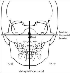
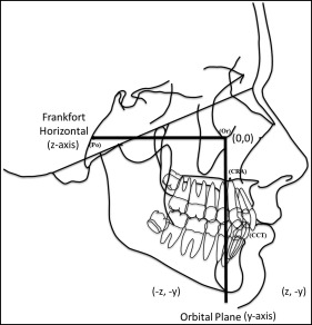
The landmarks identified on the lateral cephalogram were canine cusp tip, canine root apex, orbitale, and porion. The landmarks identified on the posteroanterior cephalogram were maxillary right canine cusp tip, maxillary right canine root apex, maxillary left canine cusp tip, maxillary left canine root apex, right orbitale, left orbitale, right ear rod, and left ear rod.
Maxillary right and left canine cusp tips and apices were identified, and the x- and y-coordinates were recorded on the posteroanterior cephalogram. The z- and y-coordinates were recorded on the lateral cephalograms of the canine cusp tip and the root apex. The z- and y-coordinates from the lateral tracing were applied to the right and left canines even though only the right canine is drawn on the Bolton standard template.
The experimental sample was composed of the posttreatment CBCT scans of 48 boys and 48 girls who had received orthodontic treatment at Case Western Reserve University in Cleveland, Ohio. They were selected based on the following criteria: white ethnicity; availability of a posttreatment CBCT volume; availability of a high-quality posttreatment photograph that showed the frontal smile; age, 15 years; presence of both maxillary right and left canines; no maxillary canine substitution treatment; and no significant restorative needs after orthodontic treatment.
No considerations were given to the manner in which the subjects in the experimental sample were treated. This sample comprised subjects with complete dentitions as well as those with extractions for orthodontic purposes. The appliance prescription, type, and manufacturer were not considered. The rationales for requiring all subjects to be 15 years of age were to provide consistency and to minimize age-related esthetic variables that could influence esthetic perceptions. Additionally, 15 years is a common age for a patient to complete his or her orthodontic treatment; this would help to ensure an adequate sample size.
The axial plane was represented by the Frankfort horizontal and was composed of a plane through the right and left orbitales extending through the right and left porions. The midsagittal plane was created at a right angle to the x-axis through the anatomic facial midline determined by inspection through crista galli and confirmed through sella turcica. The frontal plane was constructed to mimic the orbital plane and generated by creating a plane perpendicular to the Frankfort horizontal through the right and left orbitales.
The maxillary canine position was quantified by digitizing the cusp tips and root apices of the right and left canines with software (3D Imaging; Dolphin Imaging & Management Solutions, Chatsworth, Calif). The digitized point’s accuracy was confirmed by visual inspection of the other planes. The coordinates were then exported to Excel (Microsoft, Redmond, Wash) according to the Cartesian coordinate system, with 0,0,0 representing the intersection of the x-, y-, and z-axes.
Basic trigonometry was used to determine the lateral and frontal angulations of the maxillary canines. With the maxillary canine representing the hypotenuse of a right triangle, the 2 remaining sides were determined by finding the differences between the horizontal and vertical points.
The Pythagorean theorem was then used to determine the length of the maxillary canine (hypotenuse) by the formula:
A 2 ( horizontal leg ) + B 2 ( vertical leg ) = C 2 ( hypotenuse )
The angle of the maxillary canine was determined by finding the inverse tangent.
The maxillary canine angulation from the lateral view was determined in the same way, with the z-axis orientated along the horizon and maintaining the y-axis as the vertical axis. The maxillary canine angulation was determined using the inverse tangent as described previously.
One method to quantify esthetics is the Q-sort procedure. The Q-sort test is a systematic approach that has a judge generate a normal distribution based on that judge’s rankings of the esthetic outcomes being evaluated. Since this test is repeated by several judges, quantitative data of the subjects in question are generated that can be used to establish the best, worst, and average esthetic outcomes. Although a relatively limited number of methods of quantifying esthetics are available, the Q-sort method of judging esthetics is considered an effective and reliable way to quantify esthetics in a group of subjects when compared with a more absolute method of quantifying esthetics, such as the visual analog scale methodology.
The corresponding posttreatment photographs of all the subjects from the CBCT sample were gathered, and the frontal smile photograph was cropped to 3 × 5 in. The pictures were converted to gray scale, and confounding anatomic factors such as the nose, eyes, and hair were cropped from the photograph. Photoshop software (Adobe Systems, San Jose, Calif) was used to remove any blemishes that could influence a rater’s perceptions. The 48 boys and 48 girls were judged separately.
Nine third-year orthodontic residents from the Department of Orthodontics of Saint Louis University in St Louis, Mo, volunteered to perform the Q-sort. They were given consent forms in accordance with Saint Louis University’s institutional review board and agreed to participate in the study. Each resident completed 2 separate Q-sorts, one of the boys and one of the girls. The following instructions were given to each judge.
From the 48 photographs, please pick the 2 best and the 2 least esthetic smiles based only on the available photographs. Set aside the 2 best and the 2 worst at separate ends of the table. From the remaining 44 photographs, please select the 4 best and the 4 least esthetic smiles and set them aside. From the remaining 36 photographs, please select the 5 best and the 5 least esthetic smiles. From the remaining 26 photographs, please select the 8 best and the 8 least esthetic smiles. The 10 photographs remaining should represent what you consider to be average-looking smiles.
Statistical analysis
Statistics were calculated with statistical software (version 20.0; IBM, Armonk, NY). Descriptive statistics of the experimental sample were determined of the coordinate and angular measurements. The x-, y-, and z-coordinates of the canine cusp tips and apices of each subject were compared with the control sample by measuring the linear distance from the control value. To accomplish this, 6 scatter plots were generated of the canine cusp tip and the root apex of the maxillary right canine. The scatter plot was orientated along the x-y, y-z, and x-z axes and consisted of the control sample and all 96 subjects from the experimental sample.
To determine the linear distance between the normal canine coordinates and experimental subjects’ coordinates, right angle trigonometry was used.
The Q-sort was analyzed by assigning a point value to each subject depending on the esthetic category in which he or she was placed. This generated a system to quantify, rank, and distribute the esthetic outcomes. Normal distributions were also generated of the linear distances of the canine cusp tip and the root apex of the maxillary right canine. Correlations were calculated between the best and worst esthetic results as well as the best and worst canine position results.
To determine the consistency of the landmark identification of the CBCT volumes, the Cronbach alpha measure of reliability was used. A Cronbach alpha greater than 0.80 is considered reliable.
Stay updated, free dental videos. Join our Telegram channel
VIDEdental - Online dental courses


