The advent of digital photography allows the practitioner to show the patient the photographs immediately, to co-diagnose, and to work with the patient chairside or in a consult room while showing the patient some simple imaging techniques, such as whitening the teeth, making the teeth look longer, and showing the effects of orthodontics or veneers to get better alignment and other factors of smile design and esthetic dentistry. This article describes recommended digital dental photographic equipment, how to produce the standard series of diagnostic dental photographs, photographic assisted diagnosis and treatment planning including a discussion of anthropometrics and cephalometrics, and digital imaging techniques.
Standard of practice is defined as the best most often used technique, method, process, or activity that is believed to be more effective at delivering a particular outcome than any other technique, method, process, and so forth. With proper processes, a desired outcome can be delivered with fewer problems and unforeseen complications in the most efficient and effective way of accomplishing a task, based on repeatable procedures that have proven themselves over time for large numbers of people. Standard of practice refers to the level of treatment that would be expected from a competent practitioner, and is readily accepted by most practitioners in that field.
Digital dental photography is the standard of care, the standard of practice, and the best practice in esthetic dentistry.
Diagnosis and treatment planning of esthetic cases requires the use of photographs that are specifically designed to give the practitioner the information required to make that diagnosis and develop the treatment plan. Just as an orthodontist would never proceed without a cephalometric radiograph or a series of photographs, so to the “cosmetic dentist” should never proceed without a standard series of dental photographs specifically designed to assist in the diagnosis and treatment planning of the case.
Three important questions must be answered: (1) what photographs are required, (2) how do we take them, and (3) how do we interpret them to do the diagnosis and treatment planning required?
The standardization of clinical photographs was first described by this author in 1979 where the importance of standardization in views, magnification ratios, procedure, and lighting was discussed to enable the practitioner to compare photographic views before and after and between practitioners all over the world as the standard.
This series has evolved to include more photographs beyond the basic series that was designed originally for orthodontic case comparison and has now grown to involve all forms of dentistry. It should be remembered that the standard series for a cosmetic dentist will be slightly different than for an orthodontist, or general dental practitioner. Each practitioner must determine what the correct number and views will be for the clinical purposes defined. The originally described standard intraoral photographic series included 2 extraoral views (face and profile views) and 5 intraoral views (retracted anterior, right and left lateral, and 2 occlusal views).
The original intent has not changed, as the usefulness of comparison of cases before and after will always be of great benefit. But as described, the uses have expanded such that every patient will have a complete set of photographic records before treatment and after treatment to defend litigation, to improve one’s level of dental skill, to use for marketing, and of course to assist in diagnosis and treatment planning.
The advent of digital photography allows the practitioner to show the patient the photographs immediately, to co-diagnose, and to work with the patient chairside or in a consult room while showing the patient some simple imaging techniques, such as whitening the teeth, making the teeth look longer, and showing the effects of orthodontics or veneers to get better alignment and other factors of smile design and esthetic dentistry.
With progress in esthetic technique and understanding, we have come to learn other very important views to help assist the practitioner in diagnosis and treatment planning. The American Academy of Cosmetic Dentistry describes their required standard series of accreditation photographs to include a facial view, 3 smile views, 3 retracted views, 3 close-up views, and 2 occlusal views ( Fig. 1 ).


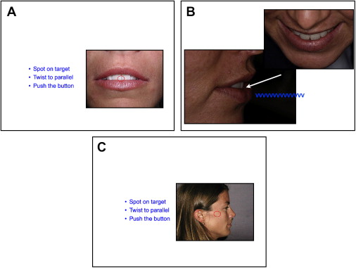
Through the work of Spear, and this author, additional views have been shown to have extreme importance in the evaluation of the maxillary incisal edge position (IEP), arguably the most important first step in the development of the new smile. These include the tooth shown at rest view, the incisal edge “VVVV” position, and the horizontal “VVVV” position to determine IEP and neutral zone horizontal position ( Fig. 2 ).
Equipment requirements and photographic principles
Dental photography equipment should be part of the dental armamentarium. It should always be available to the practitioner or staff and should therefore remain on the office premises at all times. The camera equipment you will use for digital dental photography is arguably completely different from your “at-home photography equipment.”
Photography is all about producing an image by light exposure to get a good image, and using the appropriate lens to capture that image. Digital photography captures the image onto a sensor instead of a piece of photographic film. The digital picture is then transferred by the processor in your camera to a storage card. The principles of lighting, exposure, and depth of field remain the same as with analog (film) photography.
Basic Principles
The sensitivity of the card to light is determined by the ISO. The higher the ISO, the less light is required to get a good exposure, however the higher the ISO (asa) the grainier or more pixelated the image, so in dental photography we want to use the LOWEST ISO (asa) possible for that camera, usually 100 to 200 to have a clear, crisp, sharp photograph.
The amount of light that enters the camera is controlled by the aperture, the shutter speed, and the intensity of the light.
Aperture and depth of field
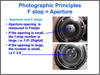
The opening in the lens called the aperture. This aperture can be changed from large to small. A large opening lets a lot of light in, whereas a small opening lets only a little bit of light in; like opening your window blinds. The opening is measured as an f:stop. The larger opening has a smaller number, ie, f:2.8, whereas a small opening has a large number, ie, f:32. The size of the opening determines the depth of field. In close-up photography everything is magnified and the depth of field is magnified also.
It is important that in dental photography everything in the photograph must be in sharp focus (high depth of field), so we always need to use the highest aperture (f:stop) possible. This means we need to have as small an opening (aperture) in the lens as is possible ( Fig. 3 ).
Shutter speed and sharpness
The next way to control the light is by the time we allow the light to enter the camera, the time the aperture stays open. This is controlled by the shutter. The longer the shutter stays open, the more light that enters, but the blurrier the photograph becomes because of camera shake and movement. A shutter speed of 1 second will allow 1 second’s worth of light to enter the camera, whereas 1/125th of a second lets only a very small amount of light to enter the camera. Because in dental photography we will always be using a flash unit (strobe) to emit the light, and because the camera and the flash synchronize with each other at a specific shutter speed (designed by each camera), usually at 1/125th of a second, this no longer becomes a variable available to us and the shutter speed is predetermined as the flash synch shutter speed.
Lighting with flash (strobe)
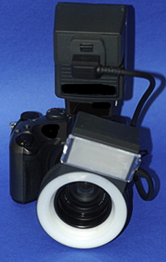
The final method is to control the intensity of the light and that is controlled by the flash unit. In regular photography the flash unit is mounted on top of the camera. In dental photography this will cast a shadow into the mouth so the flash must be mounted on the end of the lens. The flash unit can be a ring light or a point source (either will work but the ring light is easier to use). The flash should be set on its maximum intensity to allow us to use the smallest aperture (highest f:stop means highest depth of field = maximum in focus). Remember when shopping for a ring light that some lights do not have the power to illuminate the inside of the mouth using a small aperture setting at ISO of 100 to 200 ( Fig. 4 ).
The Lens
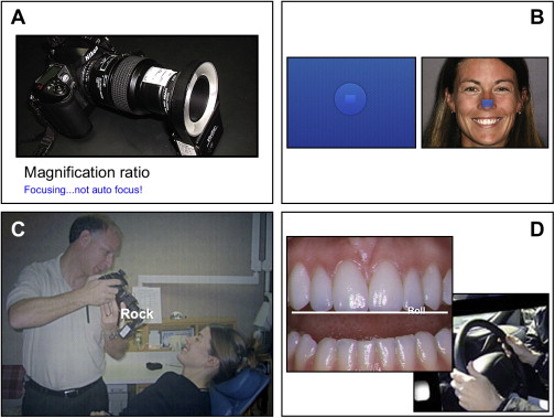
The most important piece of equipment is the lens. Macro photography is the ability to take close-up photographs. A true macro lens will give the practitioner the ability to “dial in” the magnification ratio required to take each type of photograph and get down to a 1:1 magnification ratio, which means the image in the camera is the same size as it is in real life! Watch out for the imitators here, there are lenses that are advertised as macro focus but they are not true macro lenses. Make sure the lens you purchase is a true 1:1 focusing macro lens.
A true macro lens will be able to give you a true rendition of the teeth without distortion. The focal length of the lens should be in the 90-mm to 105-mm range. The lens barrels can turn externally or internally. The external barrel will physically extend in and out. This allows you to place a piece of tape on the barrel and mark on the tape the magnification ratios, the exposure setting to help simplify the process of taking a perfect photo every time. The internal turning barrels will not extend in and out as the focusing is done internally. There will be a window in the lens to allow you to determine the magnification ratio desired ( Fig. 5 ).
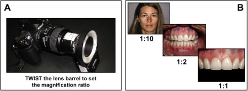
Magnification Ratio
Magnification ratio is predetermined by turning the lens barrel. In dental photography we use only 3 magnification ratios. The head shots are taken at 1:10 magnification; the smile views, occlusal views, and retracted views are taken using a 1:2 ratio; and the close-up shots are taken at a 1:1 magnification ratio. Simply turn the lens barrel and “dial in” the magnification ratio. This makes sure the view you take will be exactly the same every time.
The exposure will be the same for all the photos taken at each different magnification ratio. As you change the magnification ratio, you will then also need to change the f:stop (aperture) to correspond. This can be written down on the lens barrel itself for simplification.
Focusing
Usually when we take a photograph, we stand in one spot and push the autofocus, button which will automatically turn the lens barrel for us until the image comes into sharp focus. The problem is that this changes the magnification ratio.
In dental photography, the technique we use is a unique one. Maintaining the magnification ratio is of paramount importance, so we set the lens barrel to that magnification ratio by turning the lens barrel to that line, and instead of using the auto focus feature (turn off the auto focus feature), we focus by rocking back and forth until the image becomes clear! DO NOT TURN THE LENS BARREL TO FOCUS! (This is where a macro lens with an external barrel movement makes life easier so we can place tape on the barrel and simply turn to the correct magnification ratio.)
Camera Body
Any digital camera body that fits the lens you buy will do. The lens will be made to mount onto a particular camera, usually Nikon or Canon. Make sure the body matches the lens! Because we will be shooting everything in manual mode, we do not need to spend a lot on the camera body because we will not be using all the fancy automatic focusing features, the special automatic metering systems, and we do not even need the mega megapixels! A simple digital back that will allow you to shoot in manual mode with around 10 megapixels is more than sufficient!
SLR Versus Point and Shoot
There are varied opinions here. The costs of the systems are way down and the ease of use of the SLR system so far outweighs the point and shoot system’s needed post picture manipulation and distortion that there is no question that the SLR option is the way to go. How many gadgets do you already have in your dental closet of shame that are unused because they are just too much of a hassle to use? Using photography in your practice will create an incredible return on investment. Do not skimp on the equipment that will get the job done quickly, easily, and predictably for you and your staff.
Standard series of cosmetic photographs and how to take them
The best systems and processes in the office are repeatable, easy, and simple to follow. This allows the entire dental team to be able to efficiently produce the desired results, easily, efficiently, and perfectly every time with a minimum of training required. The trick is to understand the views you need to take. Then you need to know how to take each view using the “Goodlin Method.”
In the Goodlin Method there are 4 steps for every photograph: “Set-Spot-Rock and Roll.”
-
“Set-Spot-Rock and Roll”
- •
Set the magnification ratio and the exposure f:stop number appropriate for that view.
- •
Position the focusing point onto the spot that is desired, eg, tip of nose, papilla.
- •
Rock forward and backward to “focus.”
- •
Roll the camera like a steering wheel to make sure the image is straight.
- •
Push the button!
-
As an example, the procedure for the head shot would be as follows:
- 1.
Turn the lens barrel to 1:10 magnification ratio. The aperture will be set to about f:4 ( Fig. 6 A).

