and Angela J. Yoon1
(1)
Columbia University College of Dental Medicine and Department of Pathology and Cell Biology, Columbia University Medical Center, New York, NY, USA
A variety of papillary lesions affect the oral mucosa. In this section, we will focus on papillary lesions caused by various subtypes of human papillomavirus (HPV). HPV is a DNA virus that infects epithelial cells of the skin and mucosa resulting in solitary or multifocal epithelial lesions. Differentiating these lesions can be clinically difficult; in the following pages we present subtle clinical clues that can help. In some cases, histologic examination and HPV subtyping is needed to differentiate the lesions.
|
Papillary lesions
|
|---|
|
Verruca vulgaris
|
|
Squamous papilloma
|
|
Condyloma accuminatum
|
|
Heck disease
|
2.1 Verruca Vulgaris
Also known as the common skin wart. It is uncommon in mouth, but verruca vulgaris is very common on the skin, often occurring on the hands in children.
Clinical Appearance
Papillary, exophytic, white growth. Vary in size, most often a few millimeters to 1 cm. Multiple lesions are not uncommon. Asymptomatic.
Etiology
Viral – human papillomavirus (HPV), often as the result of auto-inoculation from cutaneous warts of the fingers. The associated HPV subtypes are that of the “low-risk” subtypes (HPV-2, HPV-4, and HPV-40).
Location
Anywhere, common in the anterior aspects of the oral cavity (i.e., anterior gingiva, lips, anterior tongue) – site that are easily auto-inoculated by putting infected fingers in mouth.
Differential Diagnosis
Squamous papilloma, condyloma acuminatum, oral lesions of Heck disease, giant cell fibroma, and verruciform xanthoma.
Treatment
Conservative surgical excision, recurrence unlikely but possible. Some lesions resolve spontaneously over time.
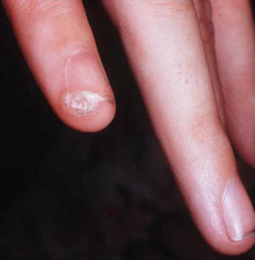
Fig. 2.1
Verruca vulagris. White warty lesion of the fingertip and nail bed of a child
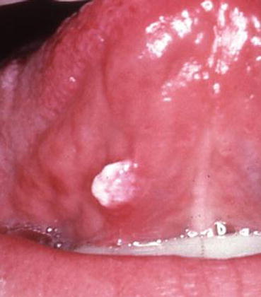
Fig. 2.2
Verruca vulagris. White, raised, papillary growth of the ventral tongue
Clinical Clue
Examining the patient’s hands for cutaneous lesion may aid in diagnosis.
2.2 Squamous Papilloma
Clinical Appearance
Papillary, exophytic, white growth, often on a stalk. Vary in size, most often a few millimeters to 1 cm. Most often lesions are solitary and asymptomatic.


