Fig. 19.1
(a) A large neuroma on the lingual nerve (arrows) at the site of damage during the removal of an impacted third molar 18 months earlier. (b) The neuroma has been excised, and the nerve ends mobilised and repaired using epineurial sutures (arrow) (From Robinson et al. [50]; reproduced with kind permission of Elsevier)
In all patients, sensation on the affected side of the tongue was assessed preoperatively and at approximately 1, 4, and 12 months or more (median: 13 months) postoperatively. Initially, each patient was asked a series of standard questions from a proforma including the following: whether the affected part of the tongue was completely numb, whether it was painful, whether they had tingling (paraesthesia) spontaneously or initiated by touching or moving the tongue, whether they tended to bite the tongue by accident, and whether they thought that their speech or taste was affected. We then made an examination to establish the presence of fungiform papillae on each side of the tongue, the presence of a palpable neuroma in the third molar region, or sensation in the tongue evoked by palpation in this region. A series of neurosensory tests was then administered by one of the authors (PPR or KGS) in a quiet room with the patient’s eyes closed and the tongue protruded. These tests included evaluation of light touch sensation using von Frey hairs, pinprick (pain) sensation, two-point discrimination, and taste sensation using both gustatory stimuli and electrogustometry. Full details of the methods used are described elsewhere [49, 50]. Finally, at the last testing session only, the patients were asked to give a subjective score to the value of the operation on a scale of 0–10. They were told that 0 indicated that the operation had been a waste of time and that 10 indicated a perfect outcome. They were told to consider this only from their own perspective, ignoring the results of the sensory testing and the perception of the surgeons.
Statistical comparisons between the results of tests at different stages were made with the chi-square test or Mann-Whitney U test as appropriate, unless otherwise stated. The effect on outcome of delay between injury and repair was assessed with Pearson’s correlation coefficient.
19.2.3 Outcome of UK Analysis
19.2.3.1 Responses to Questions
A comparison between the responses to questions asked preoperatively and at the final test is shown in Table 19.1. There was a highly significant reduction in the number of patients who thought that the affected part of the tongue was completely numb (34 to 6, p < 0.0001). A substantial proportion of the patients (n = 16) initially reported pain from the affected part of the tongue, and almost half of them (n = 25) had spontaneous paraesthesia. There was no significant change in the number reporting these problems at the final assessment, and although some patients reported a reduction in the intensity of these symptoms, we did not attempt to quantify this difference. There was a small but insignificant increase in the number of patients who reported paraesthesia initiated by touching or moving the tongue, suggesting abnormal properties of the reinnervated receptors on the tongue. Accidental tongue biting was initially a problem for 39 patients, but there was a significant reduction in this number at the final assessment (n = 26, p < 0.02). A large proportion of patients reported disturbances of speech (n = 30) and taste (n = 34), and these proportions had not changed significantly at the final assessment.
Table 19.1
A comparison between the responses to questions asked preoperatively and at the final test for patients who had undergone lingual nerve repair in the UK study
|
Preoperatively
|
Postoperatively
|
p value
|
|
|---|---|---|---|
|
Complete numbness?
|
34 (64 %)
|
6 (11 %)
|
<0.0001
|
|
Pain?
|
16 (30 %)
|
14 (26 %)
|
0.8
|
|
Spontaneous tingling?
|
25 (47 %)
|
24 (45 %)
|
1
|
|
Touch-evoked tingling?
|
9 (17 %)
|
15 (28 %)
|
0.3
|
|
Movement-evoked tingling?
|
10 (19 %)
|
17 (32 %)
|
0.2
|
|
Tongue biting?
|
39 (74 %)
|
26 (49 %)
|
<0.02
|
|
Speech affected?
|
30 (57 %)
|
30 (57 %)
|
1
|
|
Taste disturbance?
|
34 (64 %)
|
30 (57 %)
|
0.6
|
19.2.3.2 Observations
There appeared to be fewer fungiform papillae on the affected side of the tongue in 34 (74 %) of the patients preoperatively and in 24 (45 %) at the final assessment (p < 0.01). An expanded swelling, thought to be a neuroma, could be detected by palpation in five patients preoperatively and in four patients after the nerve repair operation (p = 1). Palpation in the lingual sulcus at the site where the nerve injury was expected to have occurred evoked a sensation in the tongue in 31 patients (58 %) before the operation and in 29 (55 %) after the operation.
19.2.3.3 Neurosensory Tests
On the normal, uninjured (control) side of the tongue, all patients were able to detect light touch stimuli with a 20 mN von Frey hair, pinprick stimuli of up to 150 mN, and the electrical stimuli used for electrogustometry. The two-point discrimination thresholds ranged from 2 to 10 mm (median: 4), and the ability to identify correctly the gustatory stimuli ranged from 1 to 8 (median: 6) out of eight trials.
The results of the neurosensory tests on the side of injury are shown graphically in two ways. Firstly, for the responses to light touch and pinprick stimuli, the number of patients who responded in some way to these stimuli at each assessment interval is shown in Fig. 19.2a, b. In each case, these charts show a reduction in the number of patients who responded in the early postoperative period, followed by a progressive increase beyond the initial numbers, until 42 (79 %) and 47(89 %), respectively, responded at the final test.
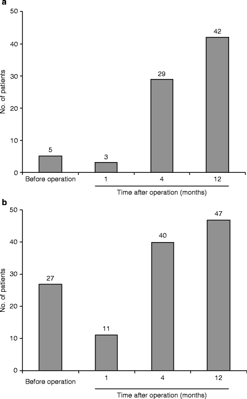
Fig. 19.2
(a) The number of patients who could detect some light touch stimuli with a 20 mN von Frey hair on the affected side, at various stages before and after operation. (b) The number of patients who could detect some pin prick stimuli of up to 150 mN on the affected side, at various stages before and after operation (From Robinson et al. [50]; reproduced with kind permission of Elsevier)
A more critical appraisal of the outcome is demonstrated by a comparison of the quantitative scores recorded at the preoperative and final postoperative assessments. These data are shown for the responses to light touch stimuli in Fig. 19.3 and indicate that 27 (51 %) of the patients responded to tests in most or all areas at the final assessment, with a highly significant improvement in the pooled responses (p < 0.0001). Equivalent data for the responses to pinprick stimuli are shown in Fig. 19.4; 41 of the patients (77 %) responded to tests in most or all areas at the final test, again with a highly significant improvement in the pooled responses (p < 0.0001). Due to the size of the tongue, two-point discrimination thresholds could be recorded for each side only if they were less than 14 mm, and the results are shown in Fig. 19.5. The pooled data again show a highly significant reduction in thresholds (p < 0.0001). Figure 19.6 indicates the extent of recovery of gustatory responses: 11 patients (21 %) could detect some taste solutions preoperatively and 33 (62 %) postoperatively (p < 0.0001). Electrogustometry evoked responses from 13 patients preoperatively, with a mean (SEM) threshold of 497 (31) μA. Postoperatively responses were evoked in 42 patients (p < 0.0001) with a mean (SEM) threshold of 312 (27) μA (p < 0.001, student’s t test).
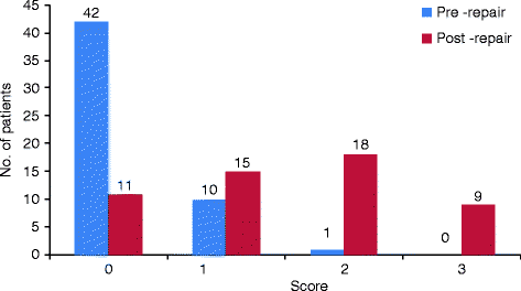
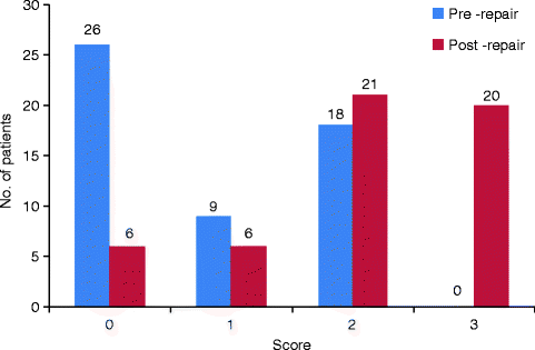
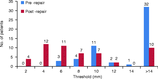
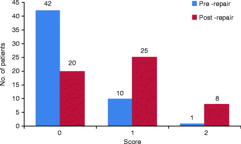
Fig. 19.3
The level of responses to light touch stimuli with a 20 mN von Frey hair, subdivided on a four-point scale: 0, no response; 1, response at the tip only; 2, response in most areas; and 3, responses apparently similar to those on the unaffected side. Preoperative data are shown in blue and responses at the final postoperative test are shown in red. The difference between the preoperative and postoperative data is highly significant (p < 0.0001)
Fig. 19.4
The level of responses to pinprick stimuli of up to 150 mN, subdivided on a four-point scale: 0, no response; 1, response at the tip only; 2, response in most areas but with an increased threshold; and 3, responses apparently similar to those on the unaffected side. Preoperative data are shown in blue, and responses at the final postoperative test are shown in red. The difference between the preoperative and postoperative data is highly significant (p < 0.0001)
Fig. 19.5
The two-point discrimination thresholds measured preoperatively (in blue) and at the final postoperative test (in red). Because of tongue size, a threshold of more than 14 mm could not be reliably assessed, and these patients have been placed in the >14 mm column. The difference between the preoperative and postoperative data is highly significant (p < 0.0001)
Fig. 19.6
The level of responses to gustatory stimuli, subdivided on a three-point scale: 0, no response; 1, some correct responses; and 2, a similar number of correct responses on each side of the tongue. Preoperative data are shown in blue, and responses at the final postoperative test are shown in red. The difference between the preoperative and postoperative data is highly significant (p < 0.0001)
19.2.3.4 Subjective Assessment
The patients’ subjective assessment of the value of the operation, taking all aspects of their experience into consideration, is shown in Fig. 19.7. The scores covered the full range from 0 to 10, with four patients reporting a score of 0 and 42 patients reporting a score of 5 or more. The median score reported by all patients was 7.
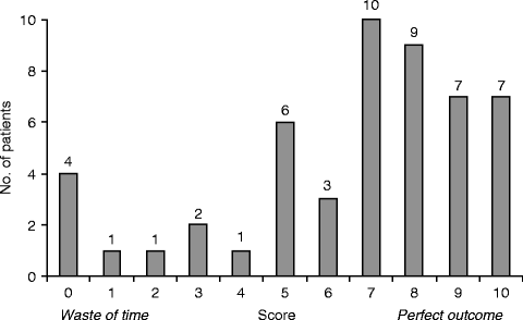
Fig. 19.7
The overall value of the operation as assessed subjectively by the patients on a scale of 0–10 (n = 51) (From Robinson et al. [50]; reproduced with kind permission of Elsevier)
19.2.3.5 Effect of Delay in Repair
The relationship between the final outcome of the nerve repair operation and the period between injury and repair was assessed for all neurosensory tests as well as the patients’ subjective scores. In no case was there any significant correlation, and an example is shown in Fig. 19.8 which shows the difference between two-point discrimination thresholds on each side of the tongue at the final test, as a good indicator of the level of recovery, plotted against the delay in repair.
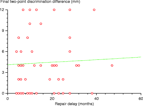
Fig. 19.8
The delay (months) between the nerve injury and repair plotted against the final outcome expressed as the difference between the two-point discrimination thresholds on the affected and unaffected sides of the tongue (p > 0.2) (From Robinson et al. [50]; reproduced with kind permission of Elsevier)
19.2.4 Patients and Methods for the Denmark Analysis
This series comprised 67 LN injuries of the patients seen in a nerve injury clinic in Copenhagen, Denmark, during the period of 1987–2005 [21]. Significantly, more patients were females (47, 70 %) than males (20, 30 %; p < 0.0001). Their median age was 30 years, with a range of 19–53 years, with no difference related to gender. The LN injuries were all caused by third molar removal, and the mean delay between injury and surgery was 12 months (range: <1–57 months).
The surgical approach was similar to that described for the UK study, except that the procedure was performed with a magnifying head loupe (3.5×) rather than an operating microscope. The neuroma on the proximal stump was resected to a level of visible fascicles, and the distal stump was cut at a level without scar tissue. From this point, a channel retractor resting on the medial cortex of the mandible was found useful to provide space for instrumentation. The nerve stumps were approximated with a holding suture if appropriate (7/0 monofilament nylon) to overcome tension, and 6–8 epineurial 8/0 monofilament nylon sutures completed the repair.
In all patients, sensation on the affected side of the tongue was assessed preoperatively and postoperatively at 1, 3, 6, and 12 months (if possible), using a standard protocol [20, 21]. Patients were questioned about sensory abnormalities (paraesthesia, dysaesthesia, etc.) and asked to rate their subjective level of sensory function on a four-point scale (0–3). In addition, the perception of tactile (feather light touch, pinprick, pointed/dull discrimination), thermal (cold, 0–20 °C and warm 45–50 °C), and location stimuli (location of touch and brush stroke direction) was assessed. Responses to each of these stimuli were rated on a four-point scale (0 = no perception of touch; 1 = perception of touch with no ability to differentiate (pointed/blunt, warm/cold, location of touch, brush stroke direction); 2 = perception with ability to differentiate less clear than normal; 3 = normal perception). The overall level of neurosensory function was characterised through the summation of all of these rating scores, therefore ranging from 0 to 21. Thus, a score of 0 signified complete absence of sensation, a score of 21 denoted normal neurosensory function, and the sensory recovery was expressed by the sum score improvement.
Two-point discrimination thresholds were assessed at 5, 10, 15, and 20 mm. Pain perception on pinching with tissue forceps was assessed through the patient’s reported experience of pain, a blink reflex, or a protective reaction, and rated as present or absent.
As soon as perception of stimuli was demonstrable, the patients were instructed to begin neurosensory retraining exercises with continued stimulation of the injured and healthy side of their tongue with recognisable items such as metal, wood, fabric, paper, and so forth.
Statistical differences between categorical scores were tested with a sign test; Chi-square or Kruskal-Wallis tests were applied to test differences between distributions.
19.2.5 Outcome of Denmark Analysis
The outcomes of nerve repair in this analysis spanned the full spectrum, with seven patients (10 %) showing no evidence of recovery, but others showing a fairly rewarding level of recovery of sensation.
19.2.5.1 Subjective Assessment of Sensation
Preoperatively, 38 patients (57 %) subjectively rated their sensory function in the affected area as 0, and the remaining 24 patients provided an average rating of 0.9 (range: 0.5–2, no data in 5 patients). Postoperatively, the subjective rating scores averaged 1.2 (range: 0–2.5).
Prior to LN repair, paraesthesia was experienced by 39 patients (58 %), 11 patients (17 %) complained of dysaesthesia, and 2 patients (3 %) had allodynia. After nerve repair, paraesthesia was still the most prevalent disturbance, felt by 40 patients (60 %), followed by 8 patients (12 %) with dysaesthesia, and 1 patient with allodynia (2 %). In 29 patients, the sensory abnormality was persistent, while in 19 it was episodic, and only 11 patients (16 %) had no neuropathic sensory disturbance (no data in 6 patients). Thus, the incidence of neurogenic discomfort was unaffected by the nerve repair.
Preoperative palpation in the lingual sulcus at the site where the nerve injury would have been expected evoked a sensation in the tongue in 55 patients (82 %), suggesting the presence of a traumatic neuroma. At the final examination, a similar response was observed in 51 patients (76 %).
19.2.5.2 Tactile Perception
Preoperative sensory testing revealed that 31 patients (46 %) had complete loss of sensation (sum score 0) on the affected side of the tongue. The remaining 36 patients had a median initial sum score of 2.0 (range 1–8), with the perception of cold being the response most frequently retained (n = 23, 34 %). After repair, the level of sensation recovered to a final median sum score of 14 (25th percentile: 9.0, 75th percentile: 16.0; min. 0, max. 20) (Fig. 19.9). The rate of recovery was fastest during the first 6 months after surgery, thereafter declining (Fig. 19.10). It appears that some patients showed a peak of sensory recovery with a subsequent loss, beyond what might have been expected from variations in neurosensory testing.
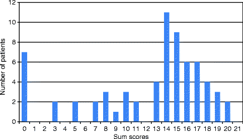
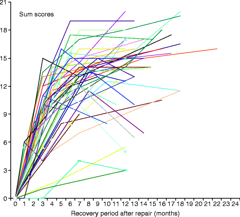
Fig. 19.9
The overall level of sensation at final follow-up examination in 65 patients after lingual nerve repair. Sum scores are the combined ratings of responses to seven test stimuli (feather light touch, pinprick, point/dull discrimination, warm, cold, location of touch, and brush stroke direction)
Fig. 19.10
The overall level of sensation (combined sum scores) at intervals after lingual nerve repair in 67 patients (From Hillerup and Stoltze [21]; reproduced with kind permission of Elsevier)
The responses to all stimuli recovered significantly, as shown in Fig. 19.11. Feather light touch and heat (45 °C) was perceived only vaguely, and the final rating score for these stimuli averaged 1 (perception of stimulus without discrimination of its quality). The remaining tests (pinprick, sharp/dull discrimination, cold, location of touch, and brush stroke direction) were generally recognised, and a score of 2 was a typical rating (perception with ability to differentiate less clear than normal). However, none of the patients regained completely normal neurosensory perception. The recovery expressed as sum score improvement correlated well with the patients subjective ratings (r = 0.58, p < 0.01).
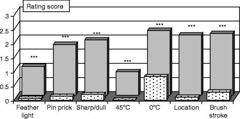
Fig. 19.11
Mean rating scores preoperatively (light shading) and at the final examination (dark shading) for responses to specific stimuli; feather light touch, pinprick, point/dull discrimination, warm, cold, location of touch, and brush stroke direction. Significant recovery for all tests, ***p < 0.001 (From Hillerup and Stoltze [21]; reproduced with kind permission of Elsevier)
19.2.5.3 Two-Point Discrimination
A two-point discrimination threshold could only be determined in one patient preoperatively (at 15 mm), but in the other patients was ≥20 mm. After nerve repair, 25 patients still had a threshold of ≥20 mm. The remaining 41 patients had a mean threshold value at the final examination of 9.9 mm. For comparison, the threshold on the uninjured side was 5.6 mm (sd. 1.8, p < 0.0001).
19.2.5.4 Pain Perception
Preoperatively, in 19 patients (28 %) pain or a pain-like sensation was perceived on pinching the affected lateral margin of the tongue. Fifty-five patients (82 %) showed pain perception at the final examination, revealing a significant recovery (p < 0.001).
19.2.5.5 Factors Influencing Recovery
When analysing the effect of age or gender on neurosensory recovery, no significant effect was found. The delay between injury and repair surgery also did not seem to influence the magnitude of recovery when considering the whole sample of 67 patients. However, assuming that other factors might be more significant determinants for poor recovery in unsuccessful cases, a test on successful cases (final sum score >9, follow-up >11 months, n = 48) was performed. This revealed that the early repair cases (operated on within the first 6 months after the injury) showed a mean improvement of sum score of 15.9 (SD 2.4, min. 12, max. 20) and thus a better recovery than patients with later repair (mean improvement of sum score – 12.6 (SD 3.1, min. 2, max. 19), p ∼ 0.002) (Fig. 19.12). The age of the patient still had no effect in this subgroup of patients (p ∼ 0.13).
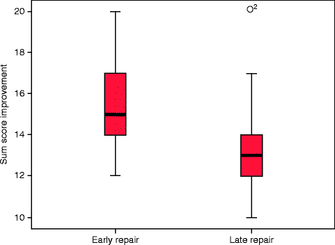
Fig. 19.12
Box plots showing improvement of sum scores in patients operated on within 6 months of injury (early repair) or after 6 months (late repair) p < 0.01. Data shown for 48 patients with a final sum score >9 (From Hillerup and Stoltze [21]; reproduced with kind permission of Elsevier)
19.2.6 Discussion on Outcome of Lingual Nerve Repair
This discussion will be structured to consider the five questions posed in the introduction.
19.2.6.1 Is Lingual Nerve Repair Worthwhile?
The extent of sensory recovery after our method of LN repair was variable, but the overall results were good. In the UK series, there were highly significant improvements in the pooled responses to light touch, pinprick, and gustatory stimuli (including electrogustometry), and highly significant reductions in the two-point discrimination thresholds. The proportion of patients who responded to most or all light touch stimuli increased from 0 to 51 % after the repair, and the proportion of patients who responded to most or all pinprick stimuli increased from 34 to 77 %. This improved level of sensation resulted in a significant reduction in the number of patients who reported accidental tongue biting, although persistent speech and taste disturbances were reported in 57 % of the patients. Most patients considered the operation worthwhile with a median subjective score of 7 on a scale of 0–10, and this equates closely with the mean score of about 2.5 on a scale of 0–4 reported for ‘global satisfaction’, by Zuniga et al. [72].
In the series of patients treated in Denmark, the outcomes were very similar. The proportion of patients with pain perception increased from 26 to 82 % after repair, the median sum score for responses to a range of seven stimuli increased from 2 to 14 (scale ranging from 0 to 21), and this correlated well with the patients’ subjective ratings. Susarla et al. [64] showed a strong correlation between neurosensory function and patient satisfaction after repair, and it is interesting to note the striking similarity between the profile of sum scores in the Denmark study, and patient subjective scores in the UK study (Figs. 19.7 and 19.9).
Comparison between our results on outcome and those reported in other studies is difficult because of variations in methods, but there are some similarities. Riediger et al. [43] indicated good or excellent recovery (with recovery of ‘protective sensitivity’) in 44 % of 16 patients who had repair by direct suture. Rutner et al. [54] reported that 90 % of their 20 patients had some improvement in neurosensory function after repair using a range of different methods. LaBanc and Gregg’s large retrospective multicentre study [27] reported that 80 % of patients could detect light touch stimuli from a von Frey hair or camel-hair brush more than 80 % of the time, but this type of study poses particular difficulties in observer consistency. A more recent retrospective review of 222 lingual nerve repairs reported complete or useful recovery of sensory function in 90.5 % of patients, although this population included injuries with a range of different aetiologies, and repaired using a range of different methods [2]. Retrospective reviews of LN repairs undertaken during two periods at the Massachusetts General Hospital reported that 81 % [65] and 75 % [13] of patients gained ‘functional sensory recovery’.
To summarise the data from the present and other studies, it seems clear that LN repair using direct reapposition by epineurial suture is a worthwhile procedure for most patients. The results seem to be better than those reported after other methods of repair such as nerve grafting [19, 36, 39], artificial conduits [41], or external neurolysis [5]. Nevertheless, the outcome of nerve repair is still not ideal as some patients do not improve, speech and taste sensation may remain affected, and recovery is rarely, if ever, complete.
19.2.6.2 Do Some Neurosensory Functions Recover Better than Others?
In the UK series, our results provided little indication of different levels of recovery for different sensory modalities. Experiments in laboratory animals have suggested this possibility, since large diameter nerve fibres seem to regenerate more successfully than small diameter fibres [9], and specific functional groups are associated with a particular range of fibre sizes. This may explain the poor recovery of the small diameter gustatory fibres in our laboratory experiments [45, 56], although it could also have occurred because few of this small population would be likely to encounter an appropriate endoneurial sheath in the distal nerve stump and be guided to a suitable taste bud receptor site. In view of this, we were slightly surprised to find the presence of some responses to gustatory stimuli in 62 % of our patients after repair. This contrasts with the results of Riediger et al. [43] who found recovery of taste sensation in only one of their patients but is consistent with the reports of Hillerup et al. [22] and Zuniga et al. [71] who reported some recovery of taste. The later report of Zuniga et al. [72] recorded return of gustatory responses in five of ten patients, which is similar to our results. The reduction in the number of patients who had fewer fungiform papillae on the side of injury after repair is a physical expression of the reinnervation by gustatory fibres and has been reported previously [48, 71].
A higher proportion of our patients responded to pinprick (painful) stimuli than light touch stimuli, in both the UK and Denmark series. It is unlikely that this indicates better regeneration of nociceptive fibres than low-threshold mechanoreceptive fibres and probably merely reflects the higher intensity of the pinprick stimuli. Similarly, in the Denmark study, the stimulus most likely to be detected after repair was a 0 °C cold stimulus, and this may have resulted from the high intensity of this stimulus rather than the preferential regeneration of cold sensitive fibres. Conversely, the Demark study found that the perception of feather light touch and warm stimuli showed the weakest recovery. Previous laboratory investigations have shown that, even after long recovery periods, reinnervated mechanoreceptors have reduced levels of sensitivity [29] and may, therefore, respond only to higher intensity stimuli. On the tongue, the reinnervated receptors may fail to respond to the 20 mN von Frey hair used in the UK study [46].
A number of other changes in the characteristics of regenerated nerve fibres have been reported in laboratory studies on both trigeminal and other peripheral nerves. These include alterations in the number and diameter of myelinated and non-myelinated axons in the proximal and distal nerve stumps [23–25], changes in conduction velocities, and alterations in mechanoreceptive fields [25, 44–46]. In view of these changes, it is not surprising that patients do not regain normal sensation after reinnervation of the tongue and continue to report some abnormalities. LaBanc and Gregg [27] noted that despite evidence of recovery, some patients still complained of ‘numbness,’ and in both the UK and Denmark studies, no patient reported completely normal sensation after nerve repair surgery.
19.2.6.3 Does Nerve Repair Surgery Reduce Dysaesthesia?
In both the UK and Denmark studies, nerve repair resulted in no significant change in the number of patients who reported pain or spontaneous paraesthesia. This is surprising since all surgeons (PPR, KGS, SH) had the impression that the symptoms of dysaesthesia were reduced by the operation. Unfortunately, neither assessment protocol included any attempt to quantify the level of these symptoms, but other studies have clearly indicated a reduction. Gregg [15] has reported a 49 % reduction in severity of pain in 31 patients after LN repair and indicated that the results seemed to vary according to the nature of the sensory disorder. Furthermore, the retrospective multicentre study by LaBanc and Gregg [27] suggested that repair resulted in a 30 % reduction in pain levels in 67.5 % of patients with hyperaesthesia. Interestingly, Pogrel and Kaban [39] reported uniformly excellent results as far as elimination of dysaesthesia was concerned, and it is difficult to explain this apparent difference in outcomes.
It has been our experience that the treatment of dysaesthesia after LN injury remains one of the most difficult clinical management problems. Pain was reported preoperatively by 30 % of the UK patients and 19 % of the Denmark patients, and the low incidence of LN dysaesthesia reported in one study is surprising [73
Stay updated, free dental videos. Join our Telegram channel
VIDEdental - Online dental courses


