Abstarct
Variations in the presence of Mandibular Foramen and Accessory Mandibular Foramen even though are rare but have been reported before. The author here reports presence of a unique Novel Aberrant Mandibular Angle foramen (NAMAF) on the lateral surface of mandibular angle which was mimicking a mandibular angle fracture.
Highlights
- •
Unusual Oval Defect Mimicking an Mandibular Angle Fracture.
- •
No Clinical Evidence of Angle fracture.
- •
Upon Exploration a Novel Foramen found on the Lateral Surface mandibular angle with a exiting neurovascular bundle.
- •
Novel Aberrant Mandibular Angle Foramen reconfirmed on CBCT postoperatively.
1
Introduction
Aberrancy in the location of the mandibular foramen as well as in the structures passing through it poses a risk for the patient undergoing any oral and maxillofacial surgical procedure [ ]. Lingual foramen [ ], accessory mental foramen [ ] and retromolar foramen [ ] are some of the frequently reported ones. A recent case report published in 2018, reported presence of bilateral coronoid foramen on the mandible [ ].
In this paper, we report presence of an unilateral foramen of its kind, Novel Aberrant Mandibular Angle foramen (NAMAF) on the lateral surface of the mandibular Angle region which was interpreted as a possible mandibular angle fracture.
Case report
A 30 year old male patient reported to the Department of Oral & Maxillofacial Surgery, Faculty of Dentistry, Jamia Millia Islamia, with the complaint of inability to chew and open his mouth accompanied with pain and discomfort. He reported with an alleged history of trauma delivered to the chin due to a road traffic accident one week back. His Clinical Examination revealed a presence of surgical dressing over a Cut Lacerated Wound (CLW) over chin, deranged occlusion with tenderness on palpation on Bilateral Temporomandibular joint (TMJ) and left mandibular parasymphseal area, a diagnosis of bilateral mandibular condylar fracture with Left mandibular symphysis fracture was made on the basis of clinical findings.
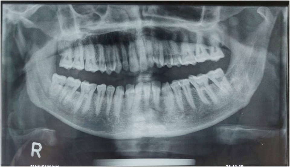
Patient was already carrying an Orthopantogram (OPG) which was advised to him by a local dispensery OPG confirmed presence of Bilateral Condylar fracture along Left parasymphseal fracture ( Fig. 1 ). Patient was advised to get a Computed Tomographic (CT) Scan of Brain and Face with mandible done, Plain CT scan of the head ruled out any injury to the brain. CT scan was performed of face with mandible in coronal, axial sections with 3-D reconstructed Images CT images revealed fracture of the bilateral mandibular condyle (Right Side Condylar Neck & Left Side Dicapitular) along with fracture at the left symphysis region. Another peculiar finding on the mandibular CT scan was presence of defect on the lateral surface of mandibular angle region ( Fig. 2 ) which suggested a possibility of mandibular angle fracture even though there were no clinical signs to support it.

The patient was advised an Open Reduction Internal Fixation (ORIF) for the mandibular fractures and was posted for same under General Anesthesia, ORIF for the Right side mandibular condylar fracture and the Left parasymphseal fracture was achieved favorably, it was decided intraoperatively to conservatively mange the Left Condylar fracture as functionally desirable occlusion was achieved by addressing above two mentioned fracture sites, It was decided to explore the left Mandibular Angle for presence of fracture. Upon reflection of the flap after intra oral incision at the left mandibular angle region, no fracture or discontinuity of the bone could be seen. Instead, a completely formed foramen with a possible neurovascular bundle exiting out of it was appreciated on the lateral surface of the left angle of mandible of ( Fig. 3 ).On carefully reexamining the coronal sections and 3-D reconstructed images of CT Scan intra operatively, an oval shape structure resembling a foramen was appreciated on few of the scan images ( Fig. 4 ) which was correlating to our clinical intraoperative finding of presence of Novel Aberrant Mandibular Angle Foramen (NAMAF) at lateral surface of angle of Mandible. Post-operative recovery of the patient was uneventful and he was stable systematically and functionally


