The nasal complex is the most frequently fractured bone in the face in the adult patient. Due to its location on the face, less force is required to fracture the nasal complex than any other facial bone. Because of the frequency of injury, as well as complex topography and different components (bone, cartilage, mucosa, skin) of the nasal complex, appropriate management of the acutely injured nose is imperative in order to prevent adverse sequelae. The purpose of this chapter is to discuss the contemporary management of bony injuries to the nasal complex.
Etiology and Pathogenesis
The most common etiologic factor for blunt nasal trauma is motor vehicle collisions followed by interpersonal injuries. The mechanism of injury is important in determining the nature of the deformity. Due to its projection, the bony complex is typically fractured in a lateral (outward) direction when the vector of the force is directly parallel to the nose. On the other hand, if the force is directed laterally to the nose (a right-handed punch to the left side of the nose), the nasal complex will then deviate away from the vector of force, with one nasal bone being medially displaced, while the other one will be laterally displaced. It is imperative to remember that the nasal septum will also be typically injured during blunt nasal trauma. The nasal septum must be appropriately reduced at the time of repair of the nasal bone; otherwise, the incidence of post-traumatic nasal deformity will significantly increase. It is also important to question the patient about the physical appearance of the nose and whether or not it is different than preinjury. Preexisting nasal bone and septal deviations should be acknowledged prior to any treatment planning.
Pathologic Anatomy
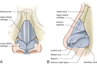
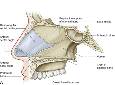
The paired nasal bones are located between the nasofrontal suture cephalically and the upper lateral cartilages caudally. They are laterally bordered by the frontal processes of the maxillary bones. It is important to remember that the nasal bones overlap the cephalic portion of the upper lateral cartilages by 3 to 4 mm ( Fig. 36-1 ). The nasal septum is composed of cartilage (anteriorly) and bone (posteriorly). The quadrangular portion of the nasal is the primary support mechanism of the nasal complex and is made of cartilage. The cartilage is thick posteriorly along the junction with the vomer and ethmoid bones, as well as along the maxillary crest ( Fig. 36-2 ). Depending on the mechanism of injury, nasal bones and septum will be displaced along specific vectors. It is just as important to properly reduce a nasal septum as it is to reduce nasal bones. As the saying goes—“as the septum goes, so goes the nose.”
Stay updated, free dental videos. Join our Telegram channel

VIDEdental - Online dental courses


