Fig. 7.1
Case I – 1 (a–h). Treatment planning: nonsurgical periodontal therapy (supra- and subgingival scaling) and conventional gingivectomy at the anterior sextant of the mandible via external beveled incision. Baseline (a). After basic procedures (b). Identification of the remaining pseudo-pockets (c, d). Pseudo-pockets surgically removed (e). One-week follow-up (f). Four-month follow-up (g). Achievement of a normal probing depth (h)

Fig. 7.2
Case II – 2 (a–c). Treatment planning: nonsurgical periodontal treatment (supra- and subgingival scaling and root planing) and frenectomy (with periosteal fenestration) at the anterior sextant. Baseline – Class III gingival recessions anterior of the mandible (a). After basic procedures – frenum surgically removed (b). Six-month follow-up – significant reduction of the gingival recession on tooth 41 (c)
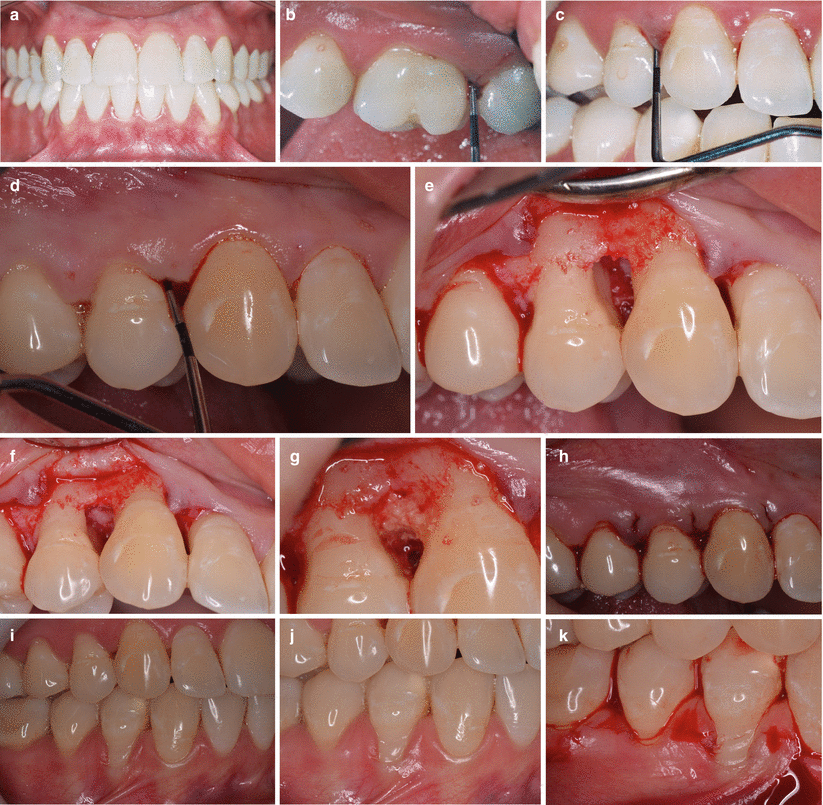
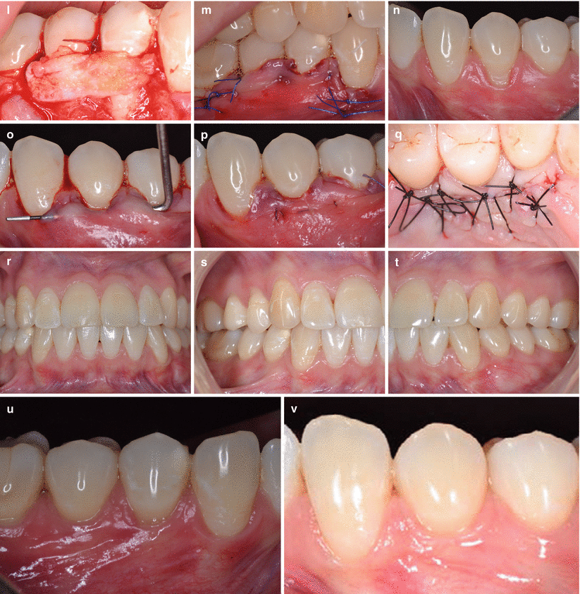
Fig. 7.3
Case III – 3 (a–v). Treatment planning: nonsurgical periodontal therapy (supra- and subgingival scaling and root planing), periodontal regeneration with bone substitute (infra-osseous defect between teeth 14 and 15), and root coverage (subepithelial connective tissue graft-based procedures) at mandibular gingival recessions. Baseline (a). Diagnosis of localized aggressive periodontitis (b, c). Presence of deep pockets (d). Infrabony defect after debridement (e). Occlusal view of the osseous defect (f). Defect – filled with bone substitute (g). Flap repositioned and sutured (h). Baseline – recession on teeth 44 and 45 (i). Baseline – closer view of the Class I and II recessions (j). Horizontal incision (k). Flap raised and graft sutured over recessions (l). Flap coronally advanced and sutured (m). Baseline – Class I gingival recession on tooth 34 (n). Tunnel flap raised (o). Graft interposed and sutured between the root surface and the tunnel flap (p). Donor site sutured (q). One-year follow-up after the last surgical procedure (r). One-year follow-up – right side (s). One-year follow-up – left side (t). One-year follow-up – teeth 44 and 45 (u). One-year follow-up – tooth 34 (v)
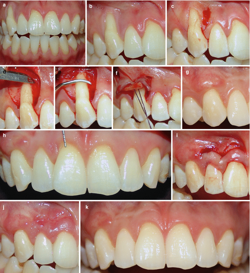
Fig. 7.4
Case IV – 4 (a–k). Treatment planning: nonsurgical periodontal therapy (supragingival scaling) and root coverage (subepithelial connective tissue graft + coronally advanced flap and modified coronally advanced flap) on the right side of the maxilla. Baseline (a). Baseline – after basic procedures (b). Realizing incisions performed adjacent to a Class II recession defect on tooth 13 (c). Flap raised by sharp dissection (d). Root planing (e). Soft tissue graft being sutured over the recession (f). Three-month follow-up (g). Shallow Class I recessions present at teeth 11 and 12 (h). Double semilunar coronally advanced flap (i). One-month follow-up (j). Six-month follow-up (tooth 13), and 3-month follow-up (teeth 11 and 12) (k)
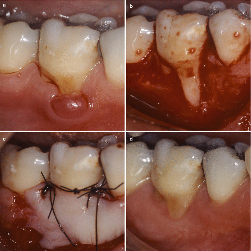
Fig. 7.5
Case V – 5 (a–d). Treatment planning: nonsurgical periodontal therapy (supragingival scaling) and removal of the pyogenic granuloma associated to a free gingival graft (*note of the authors – this lesion was diagnosed in a pregnant woman with gingivitis, and the surgical phase of treatment was conducted after the baby’s birth). Baseline – presence of a pyogenic granuloma adjacent to tooth 46 (a). Lesion removed and recipient site prepared to be grafted (b). Graft sutured to the recipient site (c). Four-month follow-up (d)
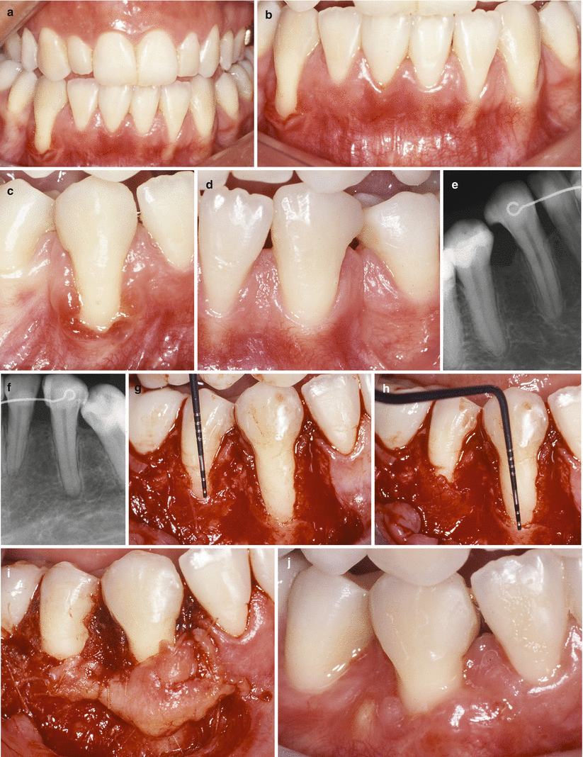
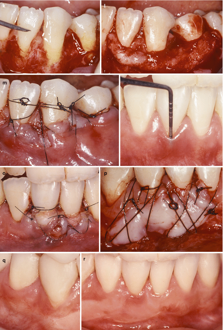
Fig. 7.6
Case VI 6 (a–r). Treatment planning: nonsurgical periodontal therapy (supra- and subgingival scaling), root coverage (subepithelial connective tissue graft + coronally advanced flap), and biotype modification (free gingival graft) at multiple sites of the mandible. Baseline (a, b). Class III gingival recession – tooth 43 (c). Class III gingival recession – tooth 33 (d). Radiographic interproximal bone loss associated to orthodontic extraction of the first mandibular bicuspids (e, f). Extension of bone dehiscence over the root surface of tooth 45 (g). Extension of bone dehiscence over the root surface of tooth 43 (h). Graft sutured over the root surface of teeth 43 and 45 (i). Two-month follow-up (j). Recipient site being prepared to accommodate the graft (k). Graft positioned (l). Graft coronally advanced and sutured (m). Baseline probing depth on tooth 41 (n). First surgical procedure at the mandibular incisors – submerged graft (o). Five months after the connective graft procedure, a free gingival graft was used to increase the thickness of keratinized tissue (p). Four-month follow-up after the last surgical procedure (q, r)
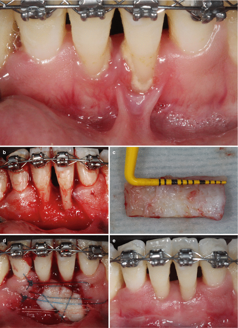
Fig. 7.7
Case VII – 7 (a–e). Treatment planning: nonsurgical periodontal therapy (supra- and subgingival scaling and root planning), frenectomy, and biotype modification concomitant orthodontic treatment. Baseline – Class IV gingival recession on teeth 31 and 41 developed during the orthodontic treatment associated to dental biofilm accumulation and high muscle insert close to the gingival margin (a). Frenum removed and osseous defect debrided (b). Length of the graft harvested from the palate (c). Graft sutured at the recipient site (d). Six-month follow-up (e)
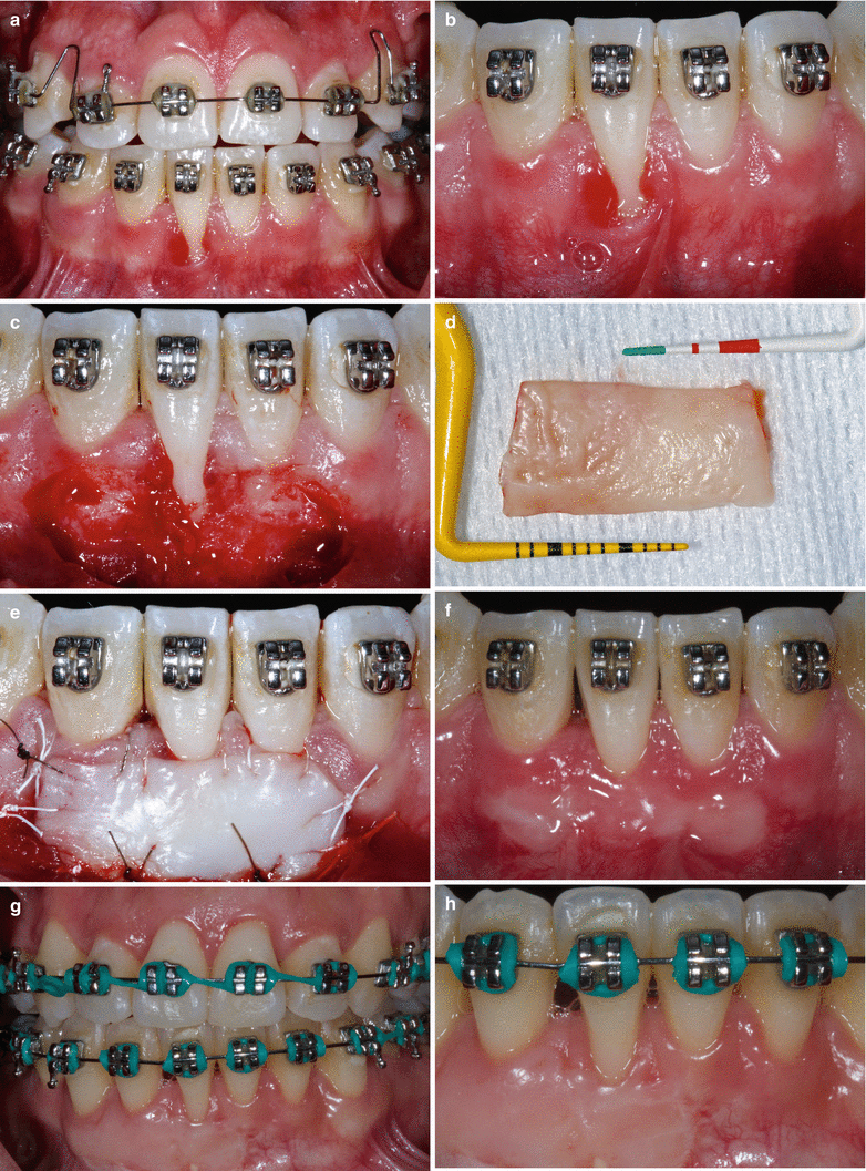
Fig. 7.8
Case VIII – 8 (a–h). Treatment planning: nonsurgical periodontal therapy (supragingival scaling), frenectomy, and biotype modification concomitant orthodontic treatment. Baseline – single Class II recession defect on tooth 41 associated to dental biofilm accumulation and a high lip frenum (a, b). Frenum and epithelial layer of the gingiva removed (c). Graft harvested from the palate (d). Graft sutured to the recipient site covering the recession (e). Three-month follow-up (f). One-year follow-up (g, h)
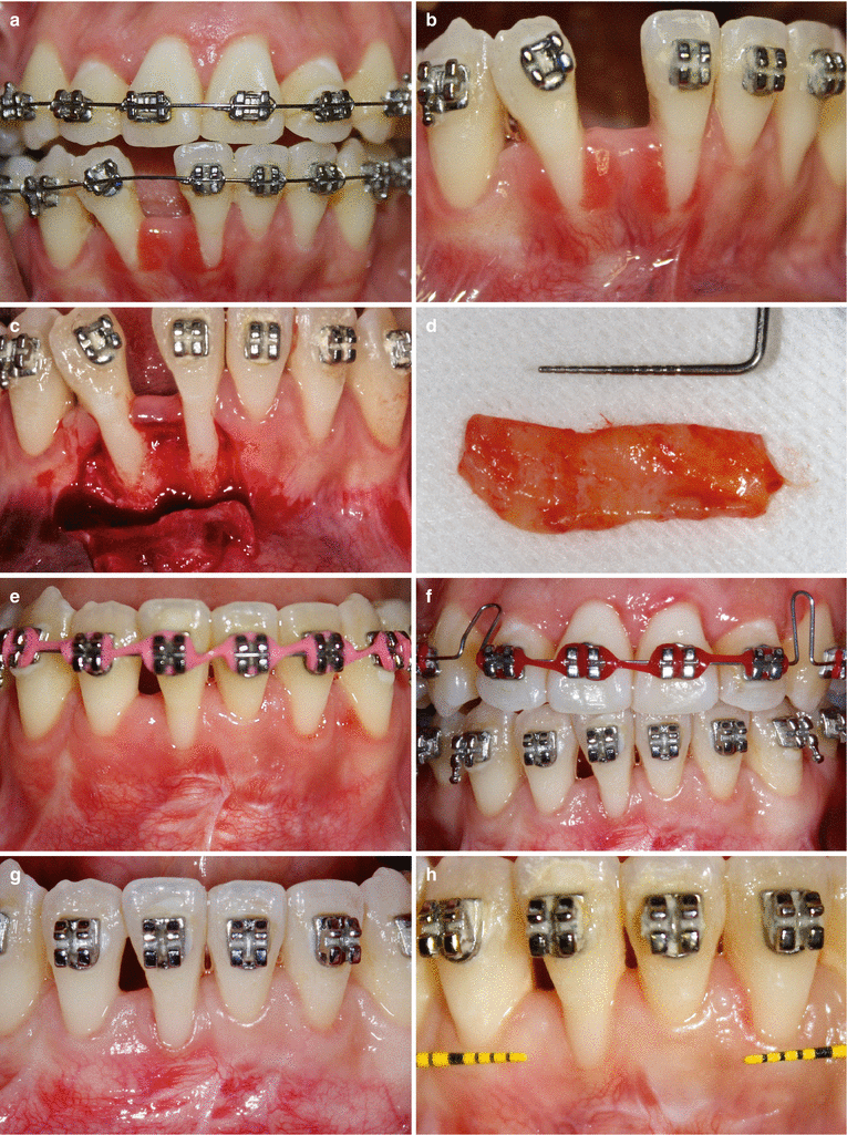
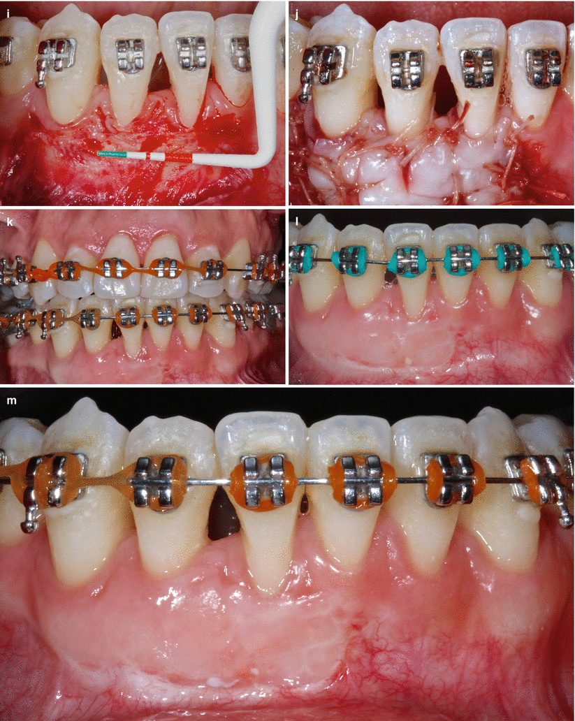
Fig. 7.9
Case IX – 9 (a–m). Treatment planning: nonsurgical periodontal therapy (supra- and subgingival scaling), root coverage (subepithelial connective tissue graft + coronally advanced flap), and biotype modification (free gingival graft) concomitant orthodontic treatment. Baseline – aggressive periodontitis patient periodontally treated and submitted to fixed orthodontics (a). Class III recession on teeth 31 and 41 associated gingival margin inflammation (b). Flap raised (c). Connective graft harvested from palate (d). Six-month follow-up (e). Six-month follow-up – baseline of the second surgical procedure (f). Baseline – second surgical procedure (g). Checking some gingival dimensions (h). Recipient site prepared to accommodate the second graft (i). Graft sutured to the recipient site (j). Three-month follow-up – second surgical procedure (k, l). Six-month follow-up – second surgical procedure (m)
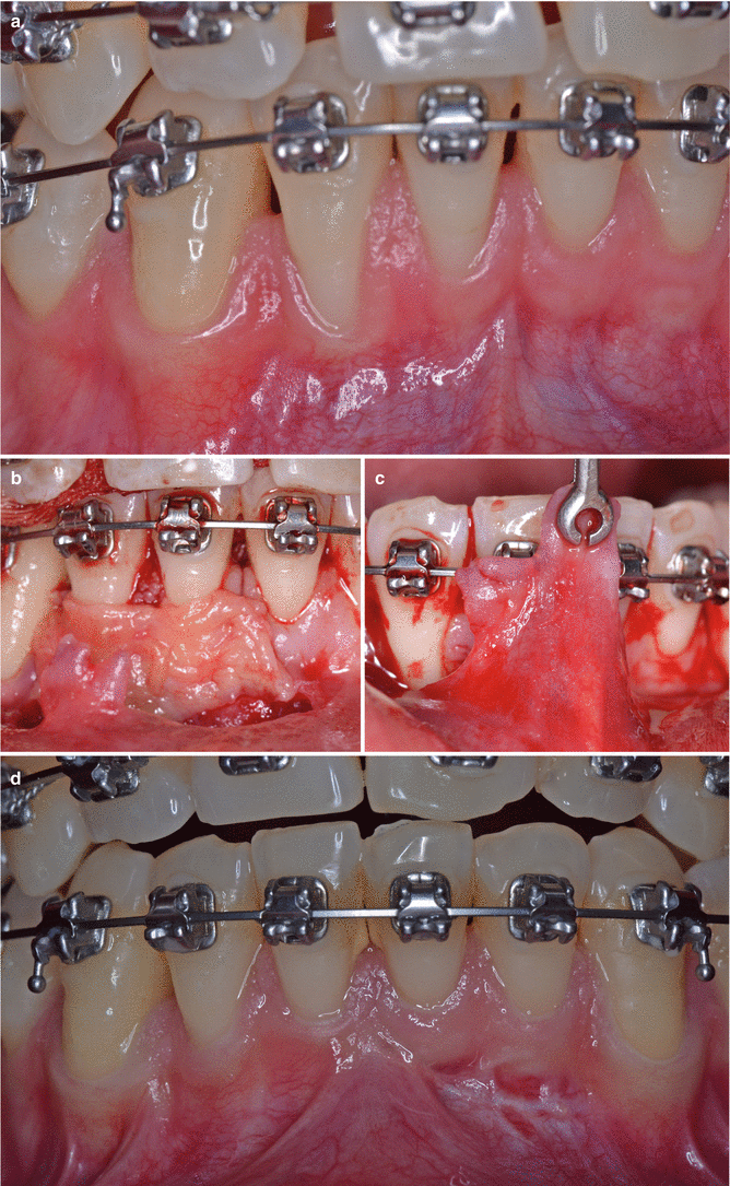
Fig. 7.10
Case X – 10 (a–d). Treatment planning: nonsurgical periodontal therapy (supragingival scaling) and root coverage (subepithelial connective tissue graft + coronally advanced flap) concomitant orthodontic treatment. Baseline – Class III gingival recessions on teeth 41 and 41 (a). Graft sutured at the recipient site (b). Checking flap tension (c). Six-month follow-up (imediately before periodontal maintenance) (d)
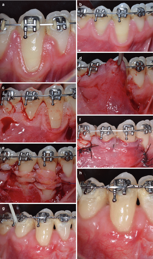
Fig. 7.11
Case X1 – 11 (a–h). Treatment planning: nonsurgical periodontal therapy (supragingival scaling) and root coverage (subepithelial connective tissue graft + coronally advanced flap) concomitant orthodontic treatment. Baseline (a). After basic procedures – Class I and II recession defects on teeth 44 and 43 (b). Horizontal and vertical incisions performed (c). Graft sutured over the recessions (d). Checking flap tension (e). Flap coronally advanced and sutured (f). One-year follow-up (g, h)
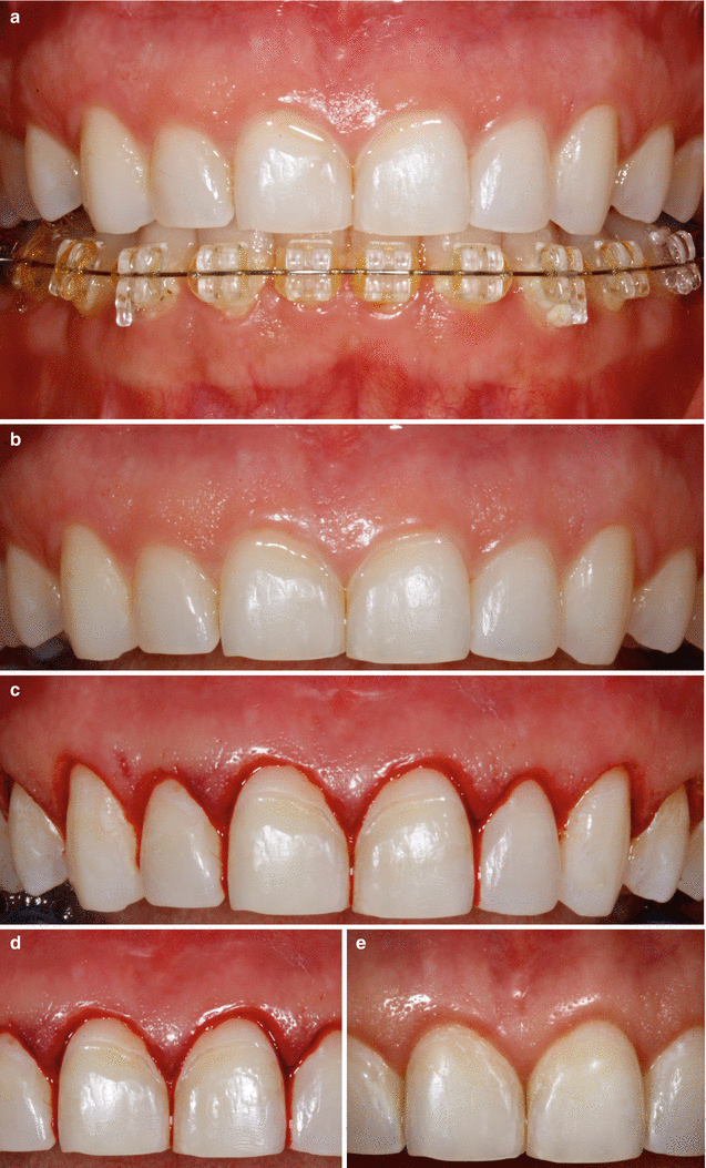
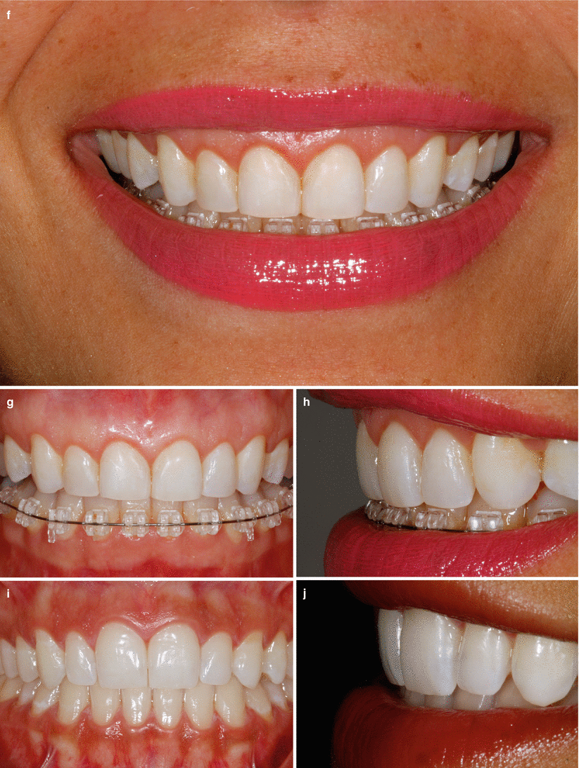
Fig. 7.12
Case XII – 12 (a–j). Treatment planning: aesthetical clinical crown lengthening of the anterior maxillary teeth after orthodontics and installation of porcelain veneers. Baseline (a, b). Gingival collar removal (c). Crown lengthening was achieved only with gingivectomy – external beveled incisions (d). Smile 3 months after surgery (e). Gingival contour around central incisors (f). Three-month follow-up (g). Three-month follow-up – lateral view (h). Six-month follow-up – porcelain veneer crowns were installed to improve anterior upper teeth’s aesthetics (i). Six-month follow-up lateral view (j)
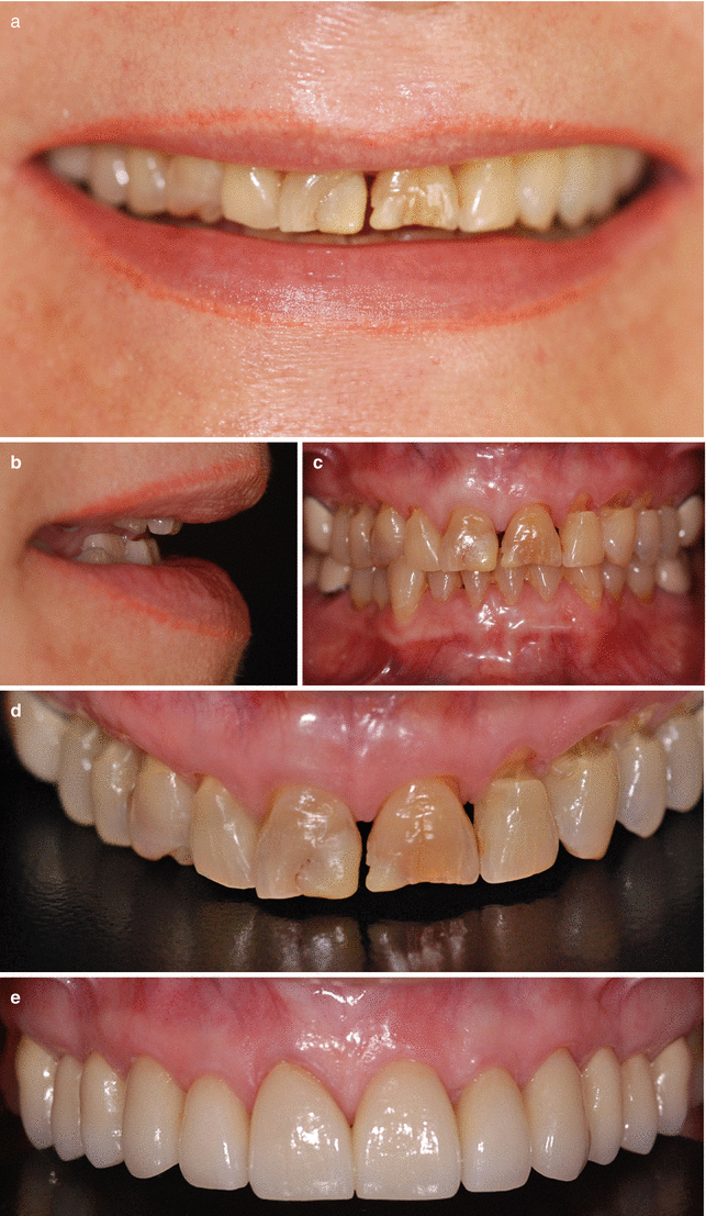
Fig. 7.13
Case XIII – 13 (a–e). Periodontal biotype modification (subepithelial connective tissue grafts) and installation of porcelain full-crown restorations. Baseline – smile (a). Baseline – lateral view (b). Baseline – clinical conditions (c, d). One year after grafting – full-crown restoration installed (e)
Stay updated, free dental videos. Join our Telegram channel

VIDEdental - Online dental courses


