Introduction
The aim of this study was to investigate the relationships between an apical curvature or a hook and the crown/root ratio in subjects with and without palatally impacted maxillary canines.
Methods
An experimental group of 44 patients (17 boys, 27 girls; mean age, 13.6 years) with 59 palatally impacted maxillary canines was selected from the records of patients referred to a radiology practice for cone-beam imaging. If a patient had bilateral palatally impacted canines, 1 canine was randomly selected for analysis. The palatally impacted canine group was matched for age and sex with 49 normal subjects (25 boys, 24 girls; mean age, 13.2 years) with 98 canines. Cone-beam DICOM files were imported into In Vivo imaging software (version 5.3; Anatomage, San Jose, Calif) for analysis. The angulations and linear variables of the maxillary canines were measured by using the software measurement tools. Chi-square and independent t tests were used to test for differences between the groups.
Results
The presence of a hook at the apical third and other root curvature were significantly different between the 2 groups ( P <0.001 and P <0.05, respectively). Of the 44 palatally impacted canines, 16 (36.4%) had an apical hook and only 1 canine in the control group had an apical hook (1.0%). The mean root length of the palatally impacted canines was 2.66 mm shorter ( P <0.001), and the mean crown/root ratio was significantly greater for the palatally impacted canines compared with the nonimpacted group ( P <0.001).
Conclusions
Palatally impacted canines have a greater tendency to develop apical hooks and are less likely to develop other root curvatures than are nonimpacted canines. Also, they have shorter roots resulting in larger crown/root ratios compared with the control group.
Highlights
- •
Apical hooks are more likely to be associated with palatally impacted canines.
- •
Hooks were frequently observed to curve in a mesial direction.
- •
Root curvature was more common in nonimpacted canines and in a distal direction.
- •
Palatally impacted canines have shorter roots and larger crown/root ratios.
The impaction of maxillary canines is a relatively common occurrence in orthodontic practice. The local etiologic factors that contribute to impaction include developmental anomalies such as malposition, shortage of space, tumors, odontomas, pericoronal pathology, and supernumerary teeth. Palatal impaction of maxillary permanent canines has also been shown to be an inherited trait and has been associated with many dental anomalies of genetic origin including small and missing teeth and short lateral incisor roots.
The term dilaceration was first coined in 1848 by Tomes, who defined the phenomenon as the forcible separation of the cap of the developed dentine from the pulp in which the development of the dentine is still progressing, and thereby creating a deviation or bend in the linear relationship of the crown of a tooth to the root (Latin: dilacero, tear up). The etiology of dilaceration is not fully understood, but it has been suggested that an acute mechanical injury to the deciduous predecessor tooth leads to the dilaceration of the underlying developing permanent tooth. However, the incidence of permanent successor tooth dilaceration is low and disproportionate to the rates of deciduous tooth injuries. Stewart in 1978 concluded that the cause of dilaceration lay in the ectopic development of the tooth germ. Furthermore, Singh and Sharma reported dilacerations among patients without a history of injury.
Many studies have been performed to investigate morphologic events associated with tooth root formation in a variety of animals, but the mechanisms involved in human tooth root formation are not well understood. Short roots, resulting in high unfavorable crown/root ratios, may affect the prognoses of teeth, especially in patients with chronic periodontitis, and may complicate orthodontic or prosthodontic treatment planning. The main reasons for short roots are disturbances during dental root development and resorption of originally well-developed roots. Underexplored topics regarding palatally impacted canines include apical dilacerations and crown/root ratios. Therefore, the aims of this study were to investigate the relationships between the presence of a severe apical curvature or a hook, other lesser root curvatures and the crown/root ratio in subjects with and without palatally impacted maxillary canines. The null hypothesis was that no relationship exists between these variables. This information could be valuable during orthodontic or surgical treatment.
Material and methods
Forty-four patients (17 boys, 27 girls) with 59 palatally impacted canines in the age range of 10 to 16 years (mean ages were 13.8 years for boys, 13.6 years for girls, and 13.6 years overall) were selected from the records of patients referred to a radiology practice for cone-beam volumetric tomography imaging. Deidentified digital imaging and communications in medicine (DICOM) files were provided for assessment. Ethics approval was granted by the Dental Sciences Research Ethics Committee, School of Dentistry, University of Queensland, in Australia. All available patients with palatally impacted canines from 10 to 16 years were included in the study. Patients with transposed palatal canines and premolars, transposed palatal canines and lateral incisors, canines with open apices, blurred images, or ongoing orthodontic treatment were excluded.
The palatally impacted canine group was matched for age and sex with 49 normal patients with 98 canines in the age range of 10 to 16 years (25 boys, 24 girls; mean ages were 13.3 years for boys, 13.1 years for girls, and 13.2 years overall); they served as the control group. A summary of the patient characteristics is shown in Table I . The subjects in the control group were provided by the same radiology practice. The DICOM data sets were produced by either Classic i-CAT (14-bit gray-scale resolution, 0.2 mm voxel size) or Next Generation i-CAT (14-bit gray-scale resolution, 0.25 mm voxel size) cone-beam 3-dimensional (3D) dental imaging systems and reconstructed with i-CAT Vision software (Imaging Sciences International, Hatfield, Pa). The DICOM files were imported into In Vivo software (version 5.3; Anatomage, San Jose, Calif) for secondary reconstruction and further investigation. The data set was reoriented according to the method of Liuk et al. Radiographic images of the canine in the coronal and sagittal planes were reconstructed from the data set, and reconstructed panoramic views were also produced.
| PIC group | Control group | |||
|---|---|---|---|---|
| Frequency | % | Frequency | % | |
| Age range (y) | ||||
| 10-10.9 | 2 | 4.5 | 3 | 6.1 |
| 11-11.9 | 3 | 6.8 | 7 | 14.3 |
| 12-12.9 | 5 | 11.4 | 8 | 16.3 |
| 13-13.9 | 10 | 22.7 | 8 | 16.3 |
| 14-14.9 | 9 | 20.4 | 6 | 12.2 |
| 15-15.9 | 8 | 18.2 | 15 | 30.6 |
| 16-16.9 | 7 | 15.9 | 2 | 4.1 |
| Sex | ||||
| Female | 27 | 61.4 | 25 | 51.0 |
| Male | 17 | 38.6 | 24 | 49.0 |
| Total | 44 | 100 | 49 | 100 |
The angulation of the apical third to the long axis of the canine root, the crown and the root lengths (along the long axis) of the palatally impacted and nonpalatally impacted canines, the locations of impacted canines (with reference to the mesiodistal position of the canine cusp tip), the vertical position of the canine cusp tip, and the angulation of the palatally impacted canine to the midsagittal plane were appraised on the volumetric images and reconstructed panoramic images using the KPG index described by Kau et al. The angular and linear measurements were made with the In Vivo software measurement tools with precision values of 0.1° and 0.01 mm, respectively.
Measurements of the canine variables were carried out as follows.
- 1.
The apical hook/curvature was measured by the deviation of the apical third of the canine root from the long axis of the canine defined by a line joining the cusp tip and the midpoint of the root, two thirds up the root. An apical hook was defined by dilaceration of the apical third of the root 50° or more to the long axis of the root. Root curvature was defined by flexion of the apical third of the root between 10° and 50° from the long axis of the tooth ( Fig 1 ).
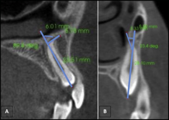
Fig 1 Method for determining whether the root was curved or had a hook: A, hook angle ≥50°; B, curvature 10° to 50°. - 2.
If the impacted canine had a hook or curvature, the direction of the hook or curvature was also determined.
- 3.
Crown length: distance between the cusp tip and the midpoint of the labial cementoenamel junction was measured.
- 4.
Root length: distance between the labial cementoenamel junction and the root apex. The root length measurements included any curvature of the root.
Statistical analysis
All measurements were performed by 1 investigator (P.V.K.S.H.). To assess intraexaminer reliability, 20 canines were randomly selected, and all measurements were repeated. These measurements were made after a reasonable time interval. The 2 sets of measurements were then compared with paired t tests. Statistical analysis was performed by using Stata software (version 12.1; StataCorp, College Station, Tex). Chi-square tests were used to analyze the significance of differences between the categorical canine variables (independent variables) and the morphology of the apical third variables (dependent variables). The measurement data were analyzed for normality by using a normal probability plot and the Shapiro-Wilks test. Since the data were normally distributed, an independent t test was used to compare the palatally impacted canine and control groups. A significance level of 0.05 was used for comparisons.
Results
The intraexaminer reliability was high, since the paired t tests indicated no significant differences ( P >0.05) between the 2 sets of measurements. The Pearson correlation coefficients were 0.55 to 0.57 ( P <0.001). Of the 44 patients with palatally impacted canines, 15 were bilateral, and 29 were unilateral.
Fifteen of the 29 unilateral impacted canines were on the right side, and 14 were on the left. Of the 15 bilateral canines, only 2 had bilateral hooks. In 1 bilateral subject, both hooks were directed labially, whereas in the other subject, 1 hook was directed labially and the other hook mesially. To avoid bias, 1 palatally impacted canine from each bilateral subject was selected randomly. Therefore, of the 59 palatally impacted canines in the study group, only 44 (23 right, 21 left) were used for the analyses.
Of the 44 palatally impacted canines, 16 (36.4%) had an apical hook, and 5 (11.4%) had curvature of the root. In the control group, only 1 canine (1.0%) had a hook, and 26 canines (26.5%) had curved roots ( Table II ). Both these variables were compared between the subjects with palatally impacted canines and the control group with a chi-square test. We found that both variables between the 2 groups were significantly different ( P <0.001 and P <0.05).
| PIC group | Control group | P value | |||||
|---|---|---|---|---|---|---|---|
| Number of teeth in boys | Number of teeth in girls | Total (of 44 teeth) | Number of teeth in boys | Number of teeth in girls | Total (of 98 teeth) | ||
| Hook | 8 | 8 | 16 (36.4%) | 0 | 1 | 1 (1.0%) | <0.001 |
| Curvature | 1 | 4 | 05 (11.4%) | 11 | 15 | 26 (26.5%) | <0.05 |
The directions of the hook and curvature of the apical third of the root for the study and control groups are shown in Table III . The most common direction of the hook in the palatally impacted canines was in the mesial direction (93.8%), and no hooks were directed distally or palatally. Only 1 hook was observed in the control group, and this was in the distal direction. Root curvature was more common in the control group (26.5%) than in the palatally impacted canine group (11.4%). Most root curvatures were directed distally. Figure 2 illustrates the different directions of the hooks in this study.


