Minor salivary gland tumors form a heterogeneous group of lesions in the head and neck because of their unique composition and ubiquitous distribution throughout the oropharynx, nasopharynx, and hypopharynx. It has been estimated that there are approximately 500 to 1000 lobules of minor salivary gland tissue dispersed throughout the oral mucosal and submucosal tissues of the lips, floor of mouth, hard and soft palates, tonsillar pillars, buccal mucosa, and tongue. These minor salivary gland lobules typically begin development in utero during the third month of gestation. As a group, salivary gland neoplasms constitute less than 3% of all head and neck tumors, but because of their remarkable structural variability, behavior, and presentation, these tumors remain an area of great clinical and research interest, while debate regarding assessment and management continues among clinicians.
Etiopathogenesis/Causative Factors
The salivary gland primordia arise as buds of proliferative epithelium from the primitive stomodeum (oral cavity) that is partly ectodermal and partly endodermal in origin, and this is responsible for the diversity seen in minor gland neoplasms. The two cells of origin for the majority of the salivary gland parenchymal neoplasms are the intercalated cell, a stem or reserve excretory cell within the salivary duct system, and the myoepithelial cell. The etiologic factors responsible for the development of salivary gland tumors are poorly understood, and only rarely there is an antecedent disease that predisposes to their development. However, controversy exists regarding the incidence of salivary gland tumors in patients with breast cancer, because studies have shown either increased incidence or no association, whereas radiation treatment primarily for benign conditions has been implicated in 11.3% of cases. Approximately 25% of all salivary gland tumors are found in the minor salivary glands, and 50% of these tumors are malignant.
Pathologic Anatomy
Usually the minor salivary glands are not clinically evident, although salivary lobules often can be palpated in the lips. For the most part, the gland lobules are 1 to 5 mm in size and separated from one another by a connective tissue stroma, but glands in the posterior hard palate tend to be more numerous and confluent. Most salivary gland lobules have individual excretory ducts that open directly into the oral cavity, but the ductal orifices are not usually visually perceptible in the normal oral mucosa. Minor salivary glands are mostly mucous type exocrine glands with the exception of those located around the circumvallate papillae and the lateral tongue that are serous in composition. The minor glands are mostly nonencapsulated and lie in close approximation with the structures around them such as the muscles of the tongue and lips. This intramuscular, nonencapsulated morphology is a factor to be considered in the histologic evaluation of invasive growth and malignancy in minor salivary gland neoplasms. Lymphatic drainage of the minor salivary gland is site specific and closely follows the lymphatic drainage of each particular site.
The glands can be grouped into vestibular, palatine, and lingual based on location in the oral cavity, and each group consists of contiguous subgroups. The vestibular glands include the labial, buccal, and retromolar subgroups, whereas the palatine group consists of the hard and soft palate and glossopalatine glands. Based on their distribution over the body of the tongue, the lingual glands are located anteriorly, posteriorly, over the faucial area, known as deep posterior groups, or within the lingual sulcus or sublingual groups.
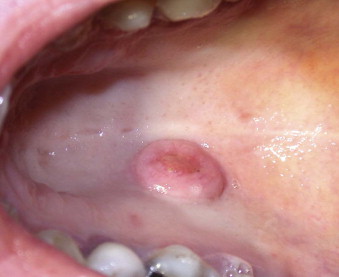
The ratio of benign to malignant neoplasms is 1.1 : 1 and most, roughly 50-60%, originate from the palatine group at the junction of the hard and the soft palates. The predominance of the pleomorphic adenoma (PA) ( Fig. 57-1 ) in the benign category and adenoid cystic carcinoma (ACC) among the malignant tumors is consistent throughout the reported case series regarding these tumors. Terminal duct adenocarcinomas and mucoepidermoid carcinomas (MECA) (intermediate or low grade) are the second most common low-grade neoplasm groups to demonstrate a strong predilection for the palate.
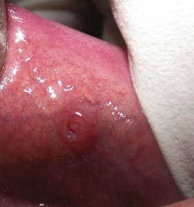
Among the vestibular glands, most benign neoplasms occur in the upper lip (84.5%) with monomorphic adenomas (inverted ductal papilloma [ Fig. 57-2 ] and sialadenoma papilliferum) occurring more commonly than pleomorphic adenomas.
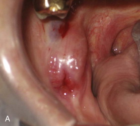
Although the lower lip is a less frequently involved site (15.5%), the majority of these neoplasms are malignant (93.8%). Once again, ACC and MECA, followed by acinic cell carcinomas, are the malignant tumors most likely to be found in this anatomic site. Although the retromolar fossa (RMF) contains only scattered minor salivary glands, it is a relatively common site of origin for MECA, which may or may not involve the underlying bone ( Fig. 57-3 ). Finally, the base of the tongue is the site of predilection for malignant minor salivary gland tumors, whereas benign tumors (which are very rare) favor the middle and anterior portions of the tongue. ACC and MECA account for roughly two thirds of the malignant neoplasms in this anatomic site. The distribution of occurrence for benign and malignant neoplasms is shown in Table 57-1 .


| SITE BENIGN | SITE MALIGNANT |
|---|---|
| Palate | Palate |
| Upper lip | Buccal mucosa/RMF |
| Buccal mucosa/RMF | Tongue |
| Lower lip | Floor of mouth |
| Tongue | Other/not specified |
| Floor of mouth | Upper lip |
| Other/not specified | Lower lip |
Pathologic Anatomy
Usually the minor salivary glands are not clinically evident, although salivary lobules often can be palpated in the lips. For the most part, the gland lobules are 1 to 5 mm in size and separated from one another by a connective tissue stroma, but glands in the posterior hard palate tend to be more numerous and confluent. Most salivary gland lobules have individual excretory ducts that open directly into the oral cavity, but the ductal orifices are not usually visually perceptible in the normal oral mucosa. Minor salivary glands are mostly mucous type exocrine glands with the exception of those located around the circumvallate papillae and the lateral tongue that are serous in composition. The minor glands are mostly nonencapsulated and lie in close approximation with the structures around them such as the muscles of the tongue and lips. This intramuscular, nonencapsulated morphology is a factor to be considered in the histologic evaluation of invasive growth and malignancy in minor salivary gland neoplasms. Lymphatic drainage of the minor salivary gland is site specific and closely follows the lymphatic drainage of each particular site.
The glands can be grouped into vestibular, palatine, and lingual based on location in the oral cavity, and each group consists of contiguous subgroups. The vestibular glands include the labial, buccal, and retromolar subgroups, whereas the palatine group consists of the hard and soft palate and glossopalatine glands. Based on their distribution over the body of the tongue, the lingual glands are located anteriorly, posteriorly, over the faucial area, known as deep posterior groups, or within the lingual sulcus or sublingual groups.
The ratio of benign to malignant neoplasms is 1.1 : 1 and most, roughly 50-60%, originate from the palatine group at the junction of the hard and the soft palates. The predominance of the pleomorphic adenoma (PA) ( Fig. 57-1 ) in the benign category and adenoid cystic carcinoma (ACC) among the malignant tumors is consistent throughout the reported case series regarding these tumors. Terminal duct adenocarcinomas and mucoepidermoid carcinomas (MECA) (intermediate or low grade) are the second most common low-grade neoplasm groups to demonstrate a strong predilection for the palate.

Among the vestibular glands, most benign neoplasms occur in the upper lip (84.5%) with monomorphic adenomas (inverted ductal papilloma [ Fig. 57-2 ] and sialadenoma papilliferum) occurring more commonly than pleomorphic adenomas.

Although the lower lip is a less frequently involved site (15.5%), the majority of these neoplasms are malignant (93.8%). Once again, ACC and MECA, followed by acinic cell carcinomas, are the malignant tumors most likely to be found in this anatomic site. Although the retromolar fossa (RMF) contains only scattered minor salivary glands, it is a relatively common site of origin for MECA, which may or may not involve the underlying bone ( Fig. 57-3 ). Finally, the base of the tongue is the site of predilection for malignant minor salivary gland tumors, whereas benign tumors (which are very rare) favor the middle and anterior portions of the tongue. ACC and MECA account for roughly two thirds of the malignant neoplasms in this anatomic site. The distribution of occurrence for benign and malignant neoplasms is shown in Table 57-1 .

| SITE BENIGN | SITE MALIGNANT |
|---|---|
| Palate | Palate |
| Upper lip | Buccal mucosa/RMF |
| Buccal mucosa/RMF | Tongue |
| Lower lip | Floor of mouth |
| Tongue | Other/not specified |
| Floor of mouth | Upper lip |
| Other/not specified | Lower lip |
Diagnostic Studies
Various diagnostic techniques—including sialography, radionucleotide scanning, ultrasonography, computed tomography scanning, and magnetic resonance imaging—are available as adjunctive studies useful in the assessment of salivary gland disorders. Most of these modalities are useful only for the major salivary glands, specifically the parotid and submandibular glands. Computed tomography (CT) and magnetic resonance imaging (MRI) are the preferred diagnostic techniques for evaluation of minor salivary gland pathology. A CT scan with contrast is easily obtained, is cost effective, and may provide valuable information regarding potential bony involvement and differentiation between inflammatory and neoplastic processes. The limitations of CT scanning include dental restoration artifacts and the inability to evaluate for perineural invasion (PNI); therefore, MRI is considered by many clinicians to be the imaging study of choice. Since the introduction of gadolinium and the marked improvement in the resolution of the images obtained by contemporary equipment, MRI is considered equal, if not superior, to contrast-enhanced CT scans. In addition, MRI permits evaluation of PNI, which is a critical factor in treatment planning of certain neoplasms, such as adenoid cystic carcinoma.
Although studies have demonstrated a correlation between findings on CT and MRI and histopathologic findings that assist in differentiation between benign and malignant processes, a histologic tissue diagnosis is mandatory for appropriate multidisciplinary treatment planning. Fine-needle aspiration biopsy (FNAB) for cytologic examination has gained popularity with increased experience among institutions in performing as well as interpreting the FNAB, and recent studies have reported sensitivity and specificity of 84.8-93.7%, with an overall accuracy of 91.1%. FNAB is valuable in the diagnosis of salivary gland neoplasms of the major salivary glands, but it has limited to no value for minor gland pathology because of the small size of the minor glands, as well as the fact that open biopsy is easily performed via transoral access for lesions in the oral cavity and oropharynx.
Both the first (1972) and second edition (1991) of the World Health Organization’s (WHO) histologic classification of the salivary gland neoplasms are based on the histologic features observed with the conventional light microscope. Immunohistochemistry, cytophotometry, molecular and cytogenic studies, and electron microscopy are all recognized as being very useful in the identification of specific neoplasms, differentiation among a variety of salivary gland tumors, as well as an assessment of their behavioral characteristics.
Stay updated, free dental videos. Join our Telegram channel
VIDEdental - Online dental courses


