Introduction
The goal of this study was to compare the dimensions of maxillary lateral incisors in subjects with and without palatally displaced canines.
Methods
An experimental group of 40 patients with 46 palatally displaced canines (20 in boys, 26 in girls; mean age, 13.9 years; range, 10.5-15.9 years) was selected from the records of patients referred to a radiology practice specializing in cone-beam volumetric tomography imaging. The palatally displaced canine group was age- and sex-matched with 30 normal subjects with 60 canines (26 in boys, 34 in girls; mean age, 13.8 years; range, 10.4-15.7 years). Cone-beam volumetric tomography DICOM files were imported into Dolphin Imaging software (version 11.0; Dolphin Imaging & Management Solutions, Chatsworth, Calif), and the volumetric images were reoriented with the long axis of the lateral incisor vertical and then reconstructed into images of a sagittal slice through the maxillary lateral incisors and 3 axial slices across the maxillary lateral incisor root. The linear variables of the maxillary lateral incisors were measured by using digital measurement tools. The widths of the maxillary lateral incisor roots were registered at the cementoenamel junction level, 4 mm apical to the cementoenamel junction level and 8 mm apical to the cementoenamel junction level. An independent t test was used to test for differences between the groups, because the data were normally distributed.
Results
In the group with palatally displaced canines, the mean length of the maxillary lateral incisors was 2.1 mm shorter ( P <0.001), and the mean root width was smaller, especially in the buccolingual dimension, by 0.7 mm ( P <0.001).
Conclusions
Lateral incisors adjacent to palatally displaced canines were smaller compared with those adjacent to normal canines.
Palatally displaced canines have been associated with lateral-incisor and other tooth anomalies. It was reported that, in an Israeli population, the prevalence of lateral incisor anomalies was 7.1%, and the prevalence values of small, peg-shaped, and missing lateral incisors were 4%, 1.8%, and 1.3%, respectively. In 1 study, 42.6% of palatally displaced canines were found to be associated with lateral incisor anomalies, 25.3% of palatally displaced canines were adjacent to small lateral incisors, 13.3% of the subjects had peg-shaped lateral incisors, and 4% of the subjects had missing lateral incisors. A meta-analysis showed that the prevalence values of congenital absence of maxillary lateral incisors were 1.55% for males and 1.78% for females.
In another study, palatally displaced canines were reported to be associated with small, peg-shaped, or missing maxillary lateral incisors, other impacted and missing teeth, and deepbite with retroclined maxillary incisors. Becker and Chaushu found that approximately half of their subjects with palatally displaced canines had delayed dental development. Chaushu et al subsequently stated that there might be 2 distinct palatally displaced canine subgroups among the male subjects but not among the female subjects. In their male sample, 1 subgroup had delayed dental development, smaller teeth, and a greatly increased prevalence of lateral incisor anomalies, but not the other subgroup that was similar to the controls. Nevertheless, Oliver found that both sexes with palatally displaced canines had delayed dental development, with a familial trend of delayed dental development among their siblings.
It has been reported that the mesiodistal crown width of the incisors was smaller in a palatally displaced canines sample. Palatally displaced canines have also been shown to be associated with short lateral incisor roots; thus, it was suggested that there is a link between lateral incisor crown size and root length. No study has previously investigated tooth lengths and root widths of lateral incisors by using 3-dimensional imaging. The aims of this study were to investigate the dimensions of maxillary lateral incisors and to compare these variables between the group with palatally displaced canines and the control group. The null hypothesis of this study was that there is no difference in the morphology of lateral incisors between these 2 groups.
Material and methods
An experimental group of 40 patients with 46 palatally displaced canines (20 in boys, 26 in girls) in the age range of 10.5 to 15.9 years (mean ages were 13.9 years for boys, 13.8 years for girls, and 13.9 years overall) were selected from the records of patients referred to a radiology practice specializing in cone-beam volumetric tomography (CBVT) imaging from October 12, 2007, to March 10, 2010. The primary records included the reconstructed panoramic images and the key cross-section images. All records were de-identified before assessment by the investigator (I.W.L.) to fulfill the ethics requirement. Ethics approval was granted by the Dental Sciences Research Ethics Committee, School of Dentistry, University of Queensland in Australia.
All available patients meeting the selection criteria were included. The inclusion criteria included palatal impaction and age range of 10 to 15.9 years. Patients with buccal or midalveolar impacted canines, mandibular impacted canines only, canines with sector I impaction, canines with zone 5 vertical position, transposed canines and premolars, transposed canines and lateral incisors, missing maxillary lateral incisors, severely resorbed maxillary lateral incisors, concurrent premolar impactions, blurred images, or ongoing orthodontic treatment were excluded.
Of the records of 251 patients with impacted canines, 40 subjects fulfilled the inclusion criteria and were available. These subjects had 46 palatally displaced canines and were age- and sex-matched with 30 normal subjects with 60 canines in the age range of 10.4 to 15.7 years (26 from boys, 34 from girls; mean ages were 13.8 years for boys, 13.8 years for girls, and 13.8 years overall); they served as the control group. The normal subjects were provided by the same radiology practice. A summary of the subjects’ characteristics is in Table I .
| PDC group | Control group | |||
|---|---|---|---|---|
| Frequency | % | Frequency | % | |
| Age range (y) | ||||
| 10-10.9 | 3 | 6.5 | 2 | 3.3 |
| 11-11.9 | 2 | 4.3 | 6 | 10.0 |
| 12-12.9 | 7 | 15.2 | 6 | 10.0 |
| 13-13.9 | 9 | 19.6 | 20 | 33.3 |
| 14-14.9 | 10 | 21.7 | 12 | 20.0 |
| 15-15.9 | 15 | 32.6 | 14 | 23.3 |
| Sex | ||||
| Female | 26 | 56.5 | 34 | 56.7 |
| Male | 20 | 43.5 | 26 | 43.3 |
| Side | ||||
| Left | 23 | 50.0 | 30 | 50.0 |
| Right | 23 | 50.0 | 30 | 50.0 |
| MDSect | ||||
| Sector 1 | 0 | 60 | 100.0 | |
| Sector 2 | 8 | 17.4 | 0 | |
| Sector 3 | 5 | 10.9 | 0 | |
| Sector 4 | 33 | 71.7 | 0 | |
| VZone | ||||
| Zone 1 | 0 | 52 | 86.7 | |
| Zone 2 | 41 | 89.1 | 8 | 13.3 |
| Zone 3 | 5 | 10.9 | 0 | |
| Resorption | ||||
| None | 36 | 78.3 | 60 | 100.0 |
| Mild | 9 | 19.6 | 0 | |
| Moderate | 1 | 2.2 | 0 | |
| Total | 46 | 100 | 60 | 100 |
The primary CBVT images were produced by either Classic i-CAT (14-bit gray-scale resolution, 0.2-0.4 mm voxel size) or Next Generation i-CAT (14 bit gray-scale resolution, 0.25-mm voxel size) cone beam 3-D dental imaging system and reconstructed through i-CAT Vision software (Imaging Sciences International, Hatfield, Pa). The digital imaging and communications in medicine (DICOM) files were imported into Dolphin Imaging software (version 11.0; Dolphin Imaging & Management Solutions, Chatsworth, Calif) for secondary reconstruction and further investigation.
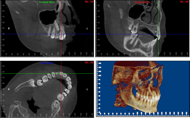
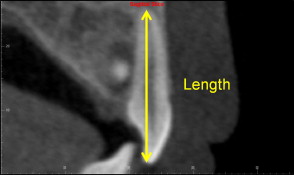
The volumetric image was reoriented with the labial surface of the lateral incisor crown facing out of the computer screen (with a tangent to the labial crown surface paralleled to the computer screen), and the lateral incisor standing vertically in the coronal and sagittal views ( Fig 1 ). This allowed a sagittal slice through the maxillary lateral incisor ( Fig 2 ) and also axial slices across the root at the cementoenamel junction (CEJ) level, 4 mm apical to the CEJ level and 8 mm apical to the CEJ level ( Fig 3 ). The thickness of each slice was set at 0.25 mm (1 voxel). The length of the lateral incisor was measured on the sagittal slice image ( Fig 2 ). The anatomically correct buccolingual and mesiodistal root widths were measured on the axial slice image across the root of the lateral incisor at the 3 levels ( Fig 3 ). The measurements were made with the Dolphin measurement tools with a precision of 0.1 mm. The images were magnified by 400% to allow a better view, yet not to produce blurred outlines or pixelated objects.