Introduction
In this prospective longitudinal study, we compared the prevalence of mandibular second molar eruption difficulties in patients treated with appliances to maintain mandibular arch perimeter. Other independent variables (age, molar angulation, space-width ratio, treatment time, and sex) were tested for their value as predictors of eruption difficulty.
Methods
Three hundred one patients and subjects were divided into 4 groups: patients treated with a Schwarz appliance, patients treated with a mandibular lingual holding arch, patients treated with a combination of both appliances, and subjects who received no treatment (controls). Logistic regression analysis was used to determine the statistical significance of the possible predictors of eruption difficulty. Panoramic radiographs were analyzed at 2 times—before and after treatment. The radiograph before treatment was evaluated for the angulation of the mandibular second molars and space available for these unerupted teeth. The radiograph after treatment was used to determine the incidence of mandibular second molar eruption difficulty.
Results
All 3 treatment groups had higher incidences of mandibular second molar eruption difficulty when compared with the controls; the increased prevalence was significant for the protocols incorporating the Schwarz appliance. Initial molar angulation, space-width ratio, age, and sex of the patient were not significant predictors of disturbances in the eruption pattern of the mandibular second molars.
Conclusions
Orthodontic appliances intended to maintain mandibular arch perimeter in the mixed dentition increase the probability of eruption disturbances of the mandibular second molars. Clinicians should monitor these patients carefully to prevent impaction of the second molars.
Nonextraction treatment often involves a therapeutic phase in the mixed dentition by means of space-maintaining or space-gaining appliances to provide a sufficient arch perimeter to align the teeth. In the mandible, the fixed lingual arch (mandibular lingual holding arch), a passive appliance, prevents the first molars from drifting forward naturally into the leeway space (about 2.5 mm per side), so that this space then can be used to alleviate crowding.
Another device aimed to preserve or increase mandibular arch length and perimeter is the Schwarz appliance. By gradual expansion, the removable mandibular Schwarz appliance serves to upright posterior teeth and also create additional arch length anteriorly. Wendling et al showed the effectiveness of the Schwarz appliance as a space maintainer for the mandibular arch in the late mixed dentition. A Schwarz appliance often is prescribed to help relieve crowding in the early or middle mixed dentition phase; a lingual arch appliance also can be used to conserve the leeway space in the late mixed dentition.
Previously published studies to date have not analyzed whether these appliances, both of which have an effect on the position of the mandibular first molars, could be causing eruption difficulties for the neighboring second molars. The prevalence rates for lack of eruption of the mandibular second permanent molar are low in the general population (about 0.05% ) and in an orthodontic population (about 1.0%). Studies on the prevalence and etiology of the eruption disturbance affecting the second permanent molar in the mandibular arch do not provide specific indications about the role of orthodontic treatment in rendering the eruption of the second molar more difficult.
This prospective controlled study was undertaken to determine the probability of mandibular second molar eruption difficulty after maintaining arch perimeter in the mixed dentition, with either the Schwarz appliance or the mandibular lingual holding arch, or a combination of the 2 appliances. The specific aims of the study also included establishing whether any specific pretreatment variables could serve as predictors of mandibular second molar eruption difficulty during orthodontic treatment aimed to preserve or increase the perimeter of the mandibular arch.
Material and methods
The treatment groups consisted of consecutively treated patients from 3 private orthodontic practices and the Graduate Orthodontic Clinic at the University of Michigan, in a large prospective study on the effects of space-maintenance appliances in the mixed dentition. The patients received treatment with the removable Schwarz appliance, the mandibular lingual holding arch, or a combination of the 2 appliances. No patients were excluded on the basis of treatment outcome.
Panoramic radiographs were taken of all treated patients at pretreatment in the mixed dentition (T1) and after treatment with the appliance and before fixed orthodontic treatment in the permanent dentition (T2).
The criteria for enrollment of patients at T1 were (1) mandibular first molars fully erupted, (2) mandibular second molars developing but not yet erupted, (3) mild to moderate crowding in the mandibular dental arch (tooth size-arch size discrepancy of 2-4.5 mm), (4) no congenitally missing or previously extracted mandibular permanent teeth (including third molars), and (5) a prepubertal stage in skeletal maturation (CS 1 or CS 2 on the basis of the cervical vertebral maturation).
The roots of the mandibular second molar had to be at least 75% formed at T2. At this developmental stage, the molar should be erupting into the oral cavity.
To estimate population variance, a pilot study was conducted. A randomly selected sample of 19 patients who were treated with the lingual arch appliance was used for the pilot study. The significance level, alpha, was set at .01, and the power, 1-beta, was set at 0.80. The hypothesized incidence was set at 1%, and the alternative incidence was set at 7%. The minimum sample size at a significance level of .01 and a power of .80 was found to be 56 patients. Therefore, enrollment was terminated when an adequate number of subjects were included in each group. Two hundred one patients participated in the trial: 85 in the lingual holding arch group, 58 in the Schwarz group, and 58 in the combination group ( Table I ).
| Group | n | Sex | Age at T1 (y) | Age at T2 (y) | |||
|---|---|---|---|---|---|---|---|
| M | F | Mean | SD | Mean | SD | ||
| S | 58 | 25 | 33 | 8.7 | 1.1 | 13.3 | 1.2 |
| L | 85 | 38 | 47 | 9.5 | 1.6 | 13.3 | 1.3 |
| SL | 58 | 30 | 28 | 8.6 | 1.1 | 13.4 | 1.1 |
| Control | 100 | 55 | 45 | 8.8 | 0.4 | 12.8 | 0.7 |
The control sample was selected from the University of Michigan Elementary and Secondary School Growth Study. Although the radiographic records from the Michigan Growth Study do not contain panoramic films, the right and left lateral oblique radiographs were considered adequate for analyzing the posterior dentition. Lateral oblique radiography has been reported to be sufficiently accurate for the identification of tooth anomalies in the posterior segment of the dental arches and for the detection of caries, when compared with panoramic radiographs. The patients were chosen based on matched age at T1 and matched dental development at T2. All Michigan Growth Study subjects were screened to fit the inclusion criteria, and the final control sample consisted of 100 patients ( Table I ).
Patients in the Schwarz group were treated with a mandibular removable Schwarz appliance. The appliance design ( Fig 1 ) involved an acrylic lingual body extending to the distal aspect of the permanent first molars. The inferior border of the acrylic lay below the gingival margin and contacted the gingival tissues. Ball clasps were incorporated into the acrylic interproximal aspect to the deciduous and permanent molars. A midline expansion screw was embedded in the acrylic lingually to the incisors. The appliance was used in patients who were in the early to middle mixed dentition.
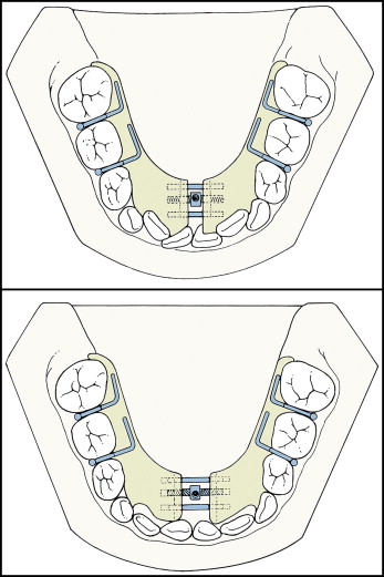
Activation involved turning the expansion screw a quarter turn per week; this produced 0.2 mm of expansion at the level of the screw. Full-time wear of the appliance was prescribed, including during meals. Depending on the severity of the lingual tipping of the posterior teeth, the activation usually continued for approximately 5 months. When activation was discontinued, the Schwarz appliance was worn as a passive retainer for at least the next 6 months. Typically, when the bonded expander was removed, the Schwarz also was discontinued, and a maxillary retention plate was delivered. No retainer was used in the mandible. The interim period, defined as the interval between the removal of the Schwarz and the date of the T2 records, on average lasted 1 year 6 months for this group.
In the lingual holding arch group, the passive mandibular lingual holding arch was constructed with bands on the mandibular first permanent molars, with a soldered .036-in stainless steel archwire extending lingually and anteriorly along the arch. Anteriorly, the archwire was fabricated to lie passively just below the cingula of the canines and the incisors ( Fig 2 ).
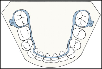
The passive lingual holding arch was used in patients who were in the late mixed dentition, before the loss of the second deciduous molars. The appliance was cemented and not removed until full eruption of the mandibular second premolars. The lingual arch was either present at the time of T2 records or removed shortly before the records were taken.
The combination group was treated first with the Schwarz appliance, according to the protocol described previously, including no mandibular retention after Schwarz appliance removal. Toward the end of the mixed dentition, the mandibular lingual holding arch was placed before the loss of the second deciduous molars. This protocol was used when, even after the Schwarz appliance had been used to gain arch length anteriorly, the maintenance of the leeway space still was helpful in obtaining sufficient arch space for the alignment of the mandibular dentition.
For the 3 treatment groups, the T2 panoramic radiographs were analyzed by 1 investigator (R.L.R.) and verified by a second investigator (J.A.M.) to determine whether an eruption difficulty existed. For the control group, the right and left lateral oblique radiographs at T2 were evaluated. The mandibular right and left second molars were placed into 1 of 2 categories.
- 1.
No eruption difficulty when, with the root formed for 75% of its length or more, the second molar had erupted in the oral cavity.
- 2.
Eruption difficulty, when the root of the mandibular second molar was at least 75% formed, but the tooth remained unerupted. This situation would require intervention to facilitate eruption of the tooth consisting of surgical exposure or uprighting of the impacted second molar.
As mentioned previously, the main purpose of this study was to evaluate the incidence of mandibular second molar eruption difficulty in different treatment groups. To focus on this objective, it was necessary to consider and control for other variables that could affect normal eruption. These variables included initial angulation of the mandibular second molar, space-width ratio, treatment time, and sex.
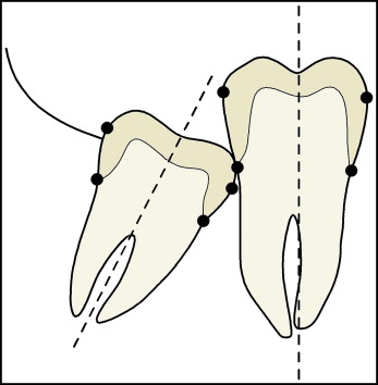
The T1 radiographs were used to analyze the initial angle formed by the long axis of the fully erupted first permanent molar and the long axis of the developing mandibular second permanent molar. The radiographs were traced by using 0.003-in frosted acetate. The long axes of these teeth were determined by a line bisecting the midpoints between the mesial and distal heights of the contours and the cementoenamel junctions, illustrated in Figure 3 . If the cementoenamel junction had not formed fully or could not be visualized adequately, the long axis was determined by constructing a line perpendicular to a line connecting the mesial and distal heights of contour on the developing crown. The angle created at the intersection of these 2 long axes was considered the initial angulation of the mandibular second molar. This analysis was performed for both the right and left sides.
For the space-width ratio, the T1 and T2 radiographs were used to determine the space available for the eruption of the mandibular second molars. To account for magnification issues as much as possible, the space-width ratio was created to determine the space available: retromolar space (mm) divided by molar width (mm) = space available for the mandibular molar on each side. If the value was ≥1, then space was adequate for the eruption of that tooth; if the value was <1, then space was inadequate space for the eruption of that tooth.
The existing retromolar space was measured from the distal surface of the first permanent molar to the intersection point of the occlusal plane and the mandibular ramus. The occlusal plane was defined as a straight line connecting the occlusal surfaces of the first molar and the fully erupted second deciduous molar. The distal surface of the first molar was indicated by a line drawn perpendicular to the occlusal plane through the distal height of contour. The ramus was traced, and the occlusal plane was extended posteriorly until this line crossed the outline of the anterior border of the ramus. The distance from the distal aspect of the first molar to the intersection of the ramus and the occlusal plane was calculated to the nearest 0.1 mm. The width of the second molar crown was also measured to the nearest 0.1 mm.
The actual times that the appliances were worn were calculated. Sex distribution was evaluated as well. All subjects in the examined groups had a postpubertal stage of skeletal maturation at T2 (CS 4 or CS 5).
To determine the accuracy of measurements taken on separate occasions by the same investigator, 10 sets of T1 radiographs were selected randomly and reanalyzed. Intraclass correlation coefficients were used to evaluate the consistency of the primary investigator (R.L.R.). Dependent t tests were also used to test for systematic errors.
Statistical analysis
By using the SPSS statistical program (version 18.0; SPSS, Chicago, Ill), means and standard deviations were calculated for the following measurements, according to the eruption difficulty category: initial angulation, space-width ratio, and treatment time. In addition, frequencies were determined for type of appliance and sex, according to the eruption difficulty category.
The data were analyzed in terms of their value as predictors of eruption difficulty. By using a statistical program (version 9.1; SAS, Cary, NC), the logistic regression analysis applied generalized estimating equations to take into account the repeated measures on each patient (ie, the correlation of the right and left sides). First, each independent variable was evaluated separately to show its effect on eruption difficulty. Then, all confounding variables were excluded to evaluate solely the effects of the different appliances. Sex distribution of the subjects with eruption difficulty was tested with the chi-square test with the Yates correction.
Stay updated, free dental videos. Join our Telegram channel
VIDEdental - Online dental courses


