Introduction
The purpose of this study was to investigate the responses of mandibular condylar cartilage to moving 2 molars in different combinations.
Methods
Rats were assigned to male and female control and experimental groups (each, n = 5). Elastic rubber bands were used to move medially the maxillary left and the mandibular right first molars in experimental group I. The same method was used to distally move the maxillary left and the mandibular right third molars, 2 mandibular third molars, and 2 maxillary third molars in experimental groups II, III, and IV, respectively. At the end of the eighth week, all condyles were examined histologically. The areas of histologic change as a percentage of total cartilage area were compared by using the Mann-Whitney U test.
Results
Cartilage degenerative remodeling was observed in experimental groups II, III, and IV. The percentage areas of degenerative remodeling were higher in female experimental groups II and III than in the female control group, and in female experimental group II than in female experimental group IV and male experimental group II (all, P <0.05).
Conclusions
The mandibular condylar cartilage of female rats responded variously to different combinations of molar movement; the most obvious remodeling was observed in groups in which the maxillary left and mandibular right third molars were moved.
Posterior teeth play a predominant role in dietary mastication, which is attributed mainly to the biomechanical action of the temporomandibular joint (TMJ). The relationship between molar occlusion and the TMJ has long been of interest. Unilateral molar lingual crossbite and non-working side interferences, as well as the loss of molar support, have been reported to be associated with specific diagnostic groups of temporomandibular disorders (TMD) or TMJ osteoarthroses. However, 2 recent reviews suggested that there is insufficient evidence to implicate occlusal factors in the etiology of TMD. Furthermore, artificial occlusal interference has been shown to exacerbate clinical signs only in subjects with a history of TMD.
Histologic studies on animals have shown that alterations in molar occlusion lead to changes in the cartilage of the TMJ, including tissue thickening ; increased tissue volume ; hyperplasia and alterations in the morphology of chondrocytes ; monocytic infiltration in condylar cartilage ; increases in chondrogenesis, osteogenesis, and angiogenesis in the posterior condyle ; and decreased proliferation of chondrocytes and the amount of extracellular matrix. However, previous animal models that investigated the relationship between abnormal molar occlusion and TMJ remodeling were all characterized by sudden changes in occlusal relationships, including the placement of an occlusal splint or metal crowns, or the unilateral removal of molars. These sudden changes in occlusion are generally different from those encountered in clinical practice.
A recent clinical study showed that a tightly locked molar occlusion, which has been suggested to be related to drifted, tilted, or supra-erupted teeth, often seen in patients who have lost posterior teeth, is associated with the incidence of TMD. A possible explanation for this observation is the altered biomechanical effect of the local contact relationship from the tightly locked occlusion on the TMJ. A supportive study on cadavers indicated that an unbalanced occlusion, including a tightly locked molar occlusion, is related to a thicker TMJ disc. Experimentally disordered occlusion in rats, achieved by moving the 2 first molars medially, was created to mimic the local contact relationship in tightly locked occlusion and was reported to have increased the thickness of the TMJ disc in the intermediate zone. However, no obvious changes in condyle cartilage were reported in that study.
Teeth are frequently moved in orthodontic therapy. A recently moved molar might not have a well-coordinated cusp-fossa relationship with its original occluding tooth. Biting with occlusal supports in various regions of the jaw has different biomechanical effects on the TMJ and the elevator muscles. Thus, it is of interest to investigate the responses of the mandibular condyle to various combinations of molar movement, which in turn cause alterations in precise cusp-fossa occlusal contact relationships in various regions of the dentition. However, to the best of our knowledge, the mandibular condylar response to moving different molars is still unknown.
In this study, we developed 4 types of experimentally disordered occlusion in rats by moving 2 different molars from their original positions. The aim of this study was to determine whether the mandibular condylar cartilage of rats responds differently to the 4 types of experimentally created disordered occlusion in terms of cartilage remodeling.
Material and methods
Twenty-five male (weight, 200-210 g) and 25 female (weight, 180-190 g) 8-week-old Sprague-Dawley rats were obtained from the animal center of the Fourth Military Medical University in Xi’an, China. All surgical procedures for and care of the animals were approved by the university ethics committee and performed according to institutional guidelines. Before the experiments, the animals were maintained for 1 week to adapt to their new environment. No obvious systemic disease or disability of movement was found in any animal. They were randomly assigned to 1 control and 4 experimental groups, each group containing 5 female and 5 male rats.
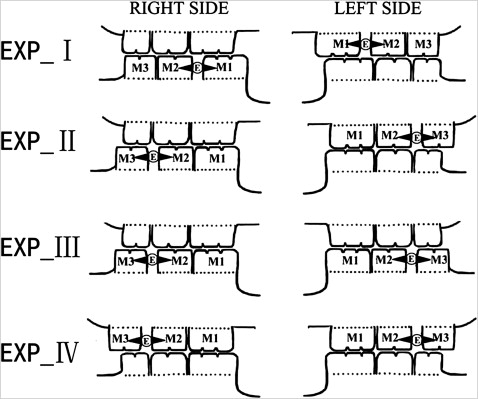
An adult Sprague-Dawley rat has 8 maxillary and 8 mandibular teeth, comprising 4 incisors and 12 molars. There is a large gap between the incisor and the first molar. For the rats in experimental group I (Exp I), an elastic rubber band (1/8 #, 3M Unitek, Monrovia, Calif), approximately 1 mm in diameter, was placed between the maxillary left first and second molars and between the mandibular right first and second molars. In this way, the first molars were moved approximately 0.8 mm medially by the elastic force of the rubber bands. One week later, the rubber band was replaced with self-curing resin (Zhangjiang Biomaterial, Shanghai, China) to maintain the gap until the end of the experiment. The same method was used to distally move the maxillary left third and the mandibular right third molars in experimental group II (Exp II), the mandibular bilateral third molars in experimental group III (Exp III), and the maxillary bilateral third molars in experimental group IV (Exp IV) ( Fig 1 ). No procedure was performed in the control group. The experimental animals and their age-matched controls were killed at the end of the eighth week of the experiment. During the experiment, no rats had any adverse signs or disturbed mastication and nutrition. All animals received the same standardized hard pellet diet, which was mainly a composite of powdered corn, wheat flour, and beans (Fourth Military Medical University, Xi’an, China).
Under deep anesthesia induced by intraperitoneal pentobarbital sodium (50 mg per kilogram of body weight), the rats were perfused through the ascending aorta with 200 mL 0.1 mol/L of phosphate buffer (pH 7.4), followed by 400 mL of 4% paraformaldehyde (4% in phosphate-buffered saline solution, pH 7.4). Tissue blocks containing the TMJs were dissected and postfixed with the same fixative overnight at 4°C and then decalcified in Kristensen’s fluid (containing sodium formate and formic acid) for 1 week. After decalcification, the tissue around the TMJ was removed, and the ramus was exposed as much as possible. The TMJs were then dehydrated in an ethanol series and embedded in paraffin wax with standardized orientation by adjusting the exposed ramus surface so that it was parallel to the upper surface of the embedding block. Serial sections, 5 μm thick, were cut through the TMJ in the sagittal plane with a microtome (RM 2135 rotary microtome, Leica Microsystems, Nussloch, Germany) and mounted on poly-l-lysine precoated glass slides. Serial sections of each condyle were stained with hematoxylin and eosin and toluidine blue for histologic assessment. To provide a reliable comparison between the areas of degraded regions in sections from different groups, the stained central sagittal sections of each condyle were selected for quantitative evaluation.
Stained histologic sections were examined under a light microscope (DM 2500, Leica, Wetzlar, Germany). Image acquisition was performed by using a Leica DFC490 system. For each section, the total areas of the condylar cartilage and the degenerative remodeling region in the cartilage were quantified by outlining the periphery of the entire cartilage and the degenerative remodeling region in the cartilage by using a computer-assisted image analyzing system (Leica QWin Plus, Cambridge, United Kingdom). Area measurements were made twice by 2 independent observers (S.-B.Y. and X.-D.L.) over 2 months, and the averaged data were used to calculate the percentages of degenerative cartilage. The percentages were then ranked for statistical analysis as follows: 0, 0%; 1, 0%-10%; 2, 10%-20%; and 3, >20%.
Statistical analysis
The ranked data were processed by using SPSS software (version 11.0, SPSS, Chicago, Ill). Since there was no significant difference ( P = 0.847) between the left and right condyles in the groups when compared with the nonparametric Wilcoxon test, the ranked data for the 2 sides were pooled and averaged for the analysis. The data for the female and male control and experimental groups (each group, n = 5) were then compared by using the nonparametric Mann-Whitney U test (α level = 0.05).
The size of the method error in measuring the areas of the entire cartilage and the degenerative remodeling region were calculated with the following formula,
method error = [ ∑ d 2 / 2 n ] ,
Stay updated, free dental videos. Join our Telegram channel
VIDEdental - Online dental courses


