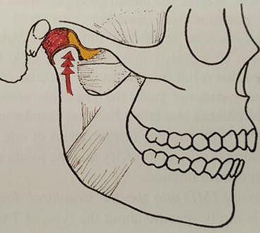
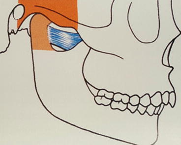
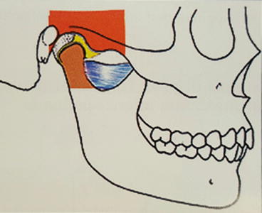
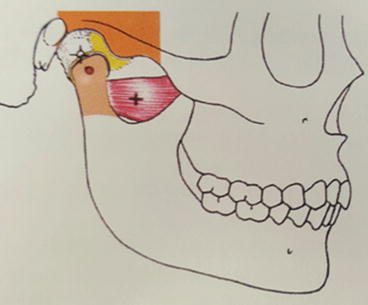
Fig. 9.1
Splayed teeth, broken incisal edge, and wear facets are indicators for a loss of vertical dimension
9.3 Types of Occlusion
Dawson classification (Dawson 2007).
Type I: Maximal intercuspation is in harmony with centric relation

-
Centric relation is variable the teeth separated
-
Treatment of TMD is not needed
-
The jaw can close to maximal intercuspation without premature contacts or deflections
-
The patient can close with no sign of discomfort
-
Use of occlusal splint is not indicated

Type IA: Maximal intercuspation occurs in harmony with centric posture

-
Intracapsular structures have deformation but have adapted
-
TMJs can accept loading with no discomfort
-
Treatment for TMD is not needed
-
Occlusal correction is not needed because there is no TMJ/occlusion disharmony

Type II: Type IIA: Condyles must displace from and adapted centric posture for maximum Intercuspation to occur

-
Discomfort from an intracapsular disorder has been ruled out.
-
The source of pain will be in the muscle or interfering teeth
-
Prognosis is excellent when occlusal interferences are eliminated
-
Surgical procedure is not indicated

Type III: Centric Relation cannot be verified

-
Treatment depends on the type of TMD and varies from a simple permissive occlusal device to relieve muscle spam to surgical procedure to correct certain types of intracapsular disorder.
-
The TMJ cannot accept load testing without discomfort.

Type IV: The occlusal relationship is in an active stage of progressive disorder because of pathological unstable TMJs.This indicate an actively progressive disorder of the TMJs that makes it impossible to stabilish a stable TMJ/occlusion relationship. Types of signs of Type IV are
-
Progressive anterior open bite
-
Progressive asymmetry
-
Progressive mandibular retrusion
-
Irreversible occlusal treatment is contra-indicated at this stage
9.4 Diagnosis of Occlusal Disorder
9.4.1 What Is “Perfected Occlusion”?
- 1.
Anterior guidance for protrusive movements. In a perfected stable occlusion, protrusive movements will be guided evenly on the central and lateral incisors. Most importantly, the cusps of the posterior teeth will never contact during this movement. There is immediate disocclusion of posterior teeth with any anterior movement, resulting in no wear on posterior teeth.
- 2.
Canine guidance for lateral movements. Lateral excursive movements will be guided on the upper and lower canine teeth. Posterior cusps should never contact during this jaw movement. Wear facets on posterior teeth are evidence that anterior guidance or canine guidance is not appropriate along with interferences involving nonworking cups. In a perfected occlusion, the posterior interferences will need to be removed.
- 3.
No CR-MIC shift (stable occlusal centric contacts. A shift indicates interferences of the posterior dentition). There should be no shift from initial centric relation contact to maximum intercuspation. For example, when watching a mandible close, if the mandible moves to either the right or left side to obtain MIC, then a shift has occurred.
9.4.2 Diagnosis: How to Determine if Occlusal Disorder Exists
- 1.
The initial exam. This is the best time to gain a thorough understanding of the stability of the patient’s TMJ. Here are some ideas of how to incorporate this evaluation into a clinical practice.
- 2.
Ask the patient if they currently have headaches or have ever suffered with headaches in the past. Are there any specific muscles that “feel tired” or sore i.e., temporalis, masseter, pterygoids, or neck muscles? Does the patient report any discomfort of their temporomandibular joint? Ask the patient to point to the sources of the pain.
Sequence of the Exam
- A.
Begin your extra oral exam by palpating the TMJ and have the patient open and close. Document any clicking, popping, crepitus, joint noises, or reported joint discomfort specific to the affected joint (refer to Chap. 1 Comprehensive Head and Neck Exams for how to do this and the form to document your findings) (Dawson 2007).

Stay updated, free dental videos. Join our Telegram channel

VIDEdental - Online dental courses


