In this chapter:
■ Success of tooth- and implant-supported restorations
■ Tooth-supported restorations
■ Esthetic complications
■ Biological complications
■ Technical complications
■ Implant-supported restorations
■ Esthetic complications
■ Biological complications
■ Technical complications
Over the years, several definitions of success have been proposed and used in restorative and implant dentistry1–3. Instead of redefining old definitions or inventing new ones over and over again, it would make more sense to move away from success definitions in general. Instead, clinicians should report whether the restorations have remained unchanged and free of any complications over the entire observation period. Hence, a “successful” restoration would be a restoration that did not require any intervention during the entire observation period4.
4.2 Success of tooth- and implant-supported restorations
In systematic reviews5,6 addressing the survival and complication rates of tooth-supported fixed dental prostheses (FDPs), only few of the included studies provided information on the number of restorations that remained intact or without complications over the observation period. The 5-year complication rate for tooth-supported FDPs was estimated to be 15.7% (95% CI: 8.5–27.7%)6,7 and for tooth-supported cantilever FDPs the respective rate was 20.6%5,7 (Table 4-1).
Table 4-1 Estimated 5-year failure and complication rates for different type of tooth-supported reconstructions and the number of abutment tooth or reconstructions analyzed

In the early days of implant dentistry the overall number of biological and technical complications was rarely reported. A former systematic review4 addressing the survival and complication rate of implant-supported FDPs could only locate three studies that gave the exact number of restorations with complications. The estimated 5-year complication rate was quite high, or 38.7% (95% CI: 33.2–44.7%)4. The most frequent complication reported in these studies was loosening of prosthetic screws. A more recent systematic review8 addressing exclusively implant-supported metal-ceramic FDPs however concluded that 15.1% (95% CI: 11.2–20.4%) of the restorations were affected by biological or technical complications over a 5-year observation period. The reduced number of complications between the older and the more recent studies might represent a positive learning curve in implant dentistry or enhanced components due to developments, causing less technical problems. Another systematic review9 addressing the survival and complication rates of metal-ceramic and zirconia-ceramic implant-supported single crowns concluded that 13.3% (95% CI: 9.0–19.3%) of the metal-ceramic and 16.2% (95% CI: 6.2–38.4%) of the zirconia-ceramic crowns experienced some kind of esthetic, biological, or technical complications over an observation period of 5 years9 (Table 4-2). Even though a significant number of studies and meta-analyses4–20 have presented impressively high survival rates for both tooth- and implant-supported restorations, it must be considered that between 15% and 20% of the restorations were affected by some kind of esthetic, biological, or technical complications. For example, a study evaluating the outcome of implant-supported restorations performed at the University of Bern21 reported a failure rate of 2.5% but an additional 16.8% of the restorations had some kind of biological and/or technical problems. Comparing the overall complication rates of tooth- and implant-supported restorations, tooth-supported restorations were more frequently affected by biological complications such as caries or loss of pulp vitality, while implant-supported restorations were more affected by technical complications such as screw loosening or material fractures.
Table 4-2 Estimated 5-year failure and complication rates for different type of implant-supported reconstructions and the number of implant abutments or reconstructions analyzed

4.3 Tooth-supported restorations
4.3.1 Esthetic complications
The incidence of esthetic complications (Fig 4-1), or restorations to be remade due to esthetic reasons, is rarely reported in the dental literature for tooth-supported single crowns (SCs) and FDPs11,12 (Table 4-1). A recent systematic review10 evaluating the outcome of tooth-supported resin-bonded fixed dental prostheses (RBFDPs) reported that only 0.3% of the included restorations had to be redone due to unacceptable esthetic appearance10. With the materials and technology available today, dental professionals should be able to imitate the natural appearance of a tooth in an acceptable way when manufacturing a tooth-supported restoration.

Fig 4-1 Central incisors with tooth-supported SCs with unacceptable esthetic outcome in a patient with a high smile line.
The current concept of how to imitate the appearance of a natural tooth with a tooth-supported restoration is presented step by step in Part I, Chapters 6 and 9.
4.3.2 Biological complications
Loss of pulp vitality
One of the most frequent biological complications affecting tooth-supported restorations is the loss of abutment tooth vitality (Fig 4-2; Table 4-1). For tooth-supported SCs, 1.8% of the abutment teeth that were considered to be vital at the time of cementation had lost vitality at an observation period of 5 years11. The loss of abutment tooth vitality was less frequent for leucite-reinforced SCs, lithium-disilicate reinforced glass-ceramic SCs, and glass-infiltrated alumina SCs compared with metalceramic and zirconia-ceramic SCs11. For tooth-supported FDPs loss of abutment tooth vitality was reported in 6.1% of the abutment teeth5,7. In the case of cantilever tooth-supported FDPs the respective number of abutment teeth with loss of vitality was 17.9% over a mean observation period of 5 years5,7. A study22 specifically addressing loss of pulp vitality in patients reconstructed with FDPs after successful treatment of advanced periodontitis, reported the highest rate of loss of abutment vitality of 8.2%. Significantly more abutment teeth lost vitality compared with non-prepared control teeth22. It must, however, be kept in mind that teeth being restored with SCs, or serving as abutments for FDPs, are often at higher risk of losing pulp vitality due to a significant amount of missing tooth substance or existing large fillings. The clinician should consider that the facial enamel-dentin thickness ranges from 1.8 mm to 3.1 mm depending on the age of the patient. The thickness also varies with tooth type and area of measurement (Fig 4-3)23. Histological changes have been detected in the pulpal tissue if the remaining dentin thickness is below 1 mm24. It is of great importance to avoid drying-out the dentin during the workflow and pulp capping in general should be avoided for abutment teeth. If endodontic treatment is needed after the restoration has been cemented then a conservative opening preparation should be implemented, leaving as much tooth substance as possible.

Fig 4-2 The pulp of tooth 22 became necrotic after trauma and a fistula can be detected on the buccal mucosa.

Fig 4-3 A histological section showing the thickness of dentin and enamel in relation to a traditional tooth preparation.
In the current concept, a minimally invasive treatment approach is recommended whenever possible. To reduce the risk of pulp necrosis, the dentin surface should be desensitized after each session. If there is doubt about the pulp vitality over time, an endodontic treatment should be considered before inserting the final restoration, respecting the age of the patient and the chance of pulpal regeneration.
Secondary caries
Another common biological complication is the development of secondary caries at the crown margins (Table 4-1). Caries has been reported at the surface25, abutment5,6,11,12,18,19, and at the restoration levels5,6,11,12,18,19. In a series of systematic reviews5,10–12 the estimated 5-year rate of secondary caries at the restoration margins was 1% for metal-ceramic SCs, 1.4% for conventional FDPs, 4.7% for cantilever FDPs, and 1.7% for RBFDPs. Most all-ceramic crowns exhibited similar 5-year caries rates as metal-ceramic SCs (Table 4-1). In contrast, the highest rate of secondary caries was reported for the first generation of densely sintered zirconia FDPs12. Some studies reported the number of restorations lost due to caries11,12. Over a 5-year observation period, 2.1% of the conventional FDPs and 1.5% of the cantilever FDPs were lost due to secondary caries (Table 4-1)5,7,12. When restoring a patient with a history of past or present caries activity (Fig 4-4), it is important to investigate the etiology of the caries first. A frequent reason for such a condition can be a reduced buffer capacity of the saliva due to general medical issues or medication influencing the salivary flow and flora. Another reason for elevated caries activity could be a patient’s dietary habits. This can usually be addressed in a motivational interview, where the patient is asked to monitor their diet in a diary for a few days. The old dogma that secondary caries can be avoided by extending crown margins subgingivally is not supported by sufficient evidence and there are even studies showing that the position of the crown margin does not influence the incidence of secondary caries26.

Fig 4-4 Old metal-ceramic restorations with visible secondary caries at several crown margins.
In the current concept a strict maintenance program is recommended for each individual patient and fluoride is applied to the root surface at every recall visit to reduce the risk of secondary caries. In case of caries prior to a treatment, its etiology should be investigated and addressed before the rehabilitation.
Recurrent periodontitis
The third biological complication is the loss of restorations due to recurrent periodontitis. Over a 5-year observation period, 1.2% of conventional tooth-supported FDPs12, 0.5% of cantilever tooth-supported FDPs5,7, and 0.8% of the RBFDPs were lost due to this complication (Table 4-1)10. It must be kept in mind that the majority of these studies were performed at known universities with excellent and strict maintenance programs5–7,10–12, which might have influenced the low rate of FDP failures due to periodontal disease. In contrast, more than 12% of the restorations were lost due to periodontal disease (Fig 4-5) in a multicenter study performed in several private practices in Saudi Arabia, most probably without regular maintenance system27–29. Today, the maintenance or periodontally supported therapy can be tailor-made for each patient using the periodontal risk assessment (PRA) tool (Figs 4-6a, b, c)30. This risk assessment tool includes: full mouth bleeding on probing index; number of residual pockets (≥ 5 mm); number of remaining teeth; ratio between bone loss evaluated on radiographs and the age of the patient; systemic or genetic factors, and environmental factors such as smoking30. The website www.perio-tools.com has all the necessary tools to plan the maintenance of periodontally compromised patients.

Fig 4-5 An old metal-ceramic restoration showing significant attachment loss around the abutment teeth on a radiograph.


Figs 4-6a to 4-6c The spiderweb – periodontal risk analysis: (a) a low-risk patient with a recommended recall interval of 12 months; (b) a medium-risk patient with a recommended recall interval of 6 months; and (c) a high-risk patient with a recommended recall interval of 3–4 months.
In the current concept, the prerequisite for the restorative phase is a successfully executed comprehensive periodontal treatment to avoid losing restorations due to recurrent periodontitis. Teeth with residual pockets (>5 mm) should not be used as abutment teeth for tooth-supported FDPs without further periodontal treatment. The crown margins are preferably placed supra- or epigingivally in periodontally compromised patients and the recall interval runs parallel to the restorative phase.
Abutment tooth fracture
Fracture of abutment teeth has been reported on the level of the abutment teeth as well as on restoration levels (Fig 4-7). For cantilever FDPs, the 5-year rate of abutment tooth fractures was 2.9%5,7. Interestingly, losing the entire restoration due to abutment tooth fracture is still a relatively rare complication as multiunit restorations can often be supported by the remaining abutment teeth if one abutment tooth fractures 5,7. In case of SCs, abutment tooth fractures were predominantly found for metal-ceramic crowns and were less frequent for all-ceramic crowns such as glass-ceramic or zirconia-ceramic crowns11,31. The estimated 5-year rate of losing a restoration due to abutment tooth fracture was reported to be 1.2% for metal-ceramic SCs, 0.9% for conventional FDPs, 1.2% for cantilever FDPs, and 0.2% for RBFDPs (Table 4-1)5,7,10–12. The risk of abutment tooth fracture is increased if the abutment tooth is endodontically treated (Fig 4-8), restored with post and core (especially if the post has a large diameter) and utilized as support for a cantilever FDP5,7,32,33.

Fig 4-7 Three endodontically treated abutment teeth of a four-unit anterior FDP fractured after 12 years in function; the restoration was lost.

Fig 4-8 A lateral incisor restored with a cast post and core and a single crown suffered a horizontal root fracture and had to be extracted.
When restoring endodontically treated teeth with a limited amount of residual tooth substance, it is important to achieve a circumferential ferrule of approximately 1.5 mm. In case of one remaining wall of dentin, a fiber-reinforced post and a composite build-up can be performed. However, if a single-rooted tooth has no remaining dentin walls, a cast post and core is preferred. If there is caries extending into the root canal, the tooth is considered irrational to treat and should be extracted.
4.3.3 Technical complications
Loss of retention
The incidence of loss of retention or fracture of the luting cement differs significantly between varying types of restorations (Table 4-1). Loss of retention is not a predominant technical problem for SCs in general. Surprisingly, zirconia-based crowns exhibited 4.7% loss of retention at 5 years11. For FDPs the 5-year rate of retention loss was on average 3.1% and also significantly higher for densely sintered zirconia FDPs compared to other material types34. The respective complication rate for conventional metal-ceramic FDPs ranged between 2.1% and 3.3%6,7,12. For cantilever FDPs, however, the 5-year rate of retention loss was significantly higher or 8.4%5,7. Loss of retention tends to correlate with increased incidence of caries, but it could not be concluded whether loss of retention or caries was the primary factor. With respect to multiunit FDPs, clinicians should investigate loss of retention carefully for each abutment tooth, since it frequently remains unnoticed by patients. As a consequence, caries can extend into a large lesion in a relatively short time.
Loss of retention can, in most cases, be avoided with proper treatment planning and appropriate preparation. With regard to traditionally cemented metal-based restorations, it is important to follow the basic principles of tooth preparation, aiming for a preparation convergence angle of approximately 10 degrees. A minimal wall height of 3 mm for small diameter teeth like premolars, canines, and incisors and a minimal wall height of 4 mm for molars should be respected35. If this can’t be achieved, additional measures such as surgical crown lengthening, build-up with post and core, or adhesive cementation should be implemented. For traditionally cemented restorations the resisting form of the preparation, which supports the restoration when being exposed to lateral forces, is as important as the retention form. Additional preparation boxes or grooves can be an option to increase the retention and resistance form of traditionally cemented restorations. If the basic principles of tooth preparation cannot be followed due to a limited amount of tooth substance, it is important to change the treatment approach and opt for adhesive cementation. The prerequisites for the adhesive cementation are the evaluation of the quality of enamel and dentin (see Part I, Chapter 9), well-fitting restorations, and to meticulously follow the recommended steps of the cementation process depending on the adhesive system. In case of digital fabrication of a restoration, the cement space should be adjusted according to the retentiveness of the tooth preparation. Hence, for an abutment tooth with short preparation walls the width of the cement space should be set at the minimum, whereas for long preparation walls the dental technician can work with a standard cement space. If considered necessary, additional surface treatment such as sandblasting can be applied to increase the roughness of the restoration.
Debonding or loss of retention (Fig 4-9) is by far the most frequent technical complication for RBFDPs with a reported annual complication rate ranging between 0% and 12.8%. The overall 5-year debonding rate was reported to be 15.0%10. The incidence of RBFDPs debonding significantly depends on the framework material used and the cementation procedure. A recent systematic review10 analyzing the outcome of RBFDPs reported an annual debonding rate of 2.9% for metal-ceramic RBFDPs, 4.2% for metal-acrylic RBFDPs, 1.7% for fiber-reinforced composite RBFDPs, and 1.4% for zirconia framework RBFDPs. However, for RBFDPs with glass-infiltrated and glass-reinforced ceramic frameworks no debonding occurred. Moreover, the incidence of debonding was higher for RBFDPs inserted in the posterior area compared with RBFDPs placed in the anterior10.


Figs 4-9a and 4-9b The patient did not notice that the metal-ceramic RBFDP lost retention at one retainer and a large caries lesion developed in few months.
Today, zirconia- or glass-reinforced ceramic such as lithium-disilicate ceramic are the favorable framework materials when manufacturing RBFDPs. The risk of fracture is less for zirconia as framework material compared with reinforced glass-ceramic.
Thus, the current concept recommends the use of zirconia, even though glass-reinforced ceramic RBFDPs show an excellent retention to enamel even without any preparation. Another advantage of zirconia is that retainers can be made thinner, which increases the indication spectrum of RBFDPs. To reduce the risk of debonding for zirconia-based RBFDPs, it is recommended to prepare two parallel retention grooves and a central stop for the retainer. In situations with minimal retention area, eg, short teeth, the dental technician can apply a microveneering of feldspar ceramic on the inside of the retainer, allowing for acid etching of the retainer.
Fracture of core or framework
Core fractures are a rare event for metal-ceramic SCs and occur in only 0.03% at 5 years11. The incidence of core fractures is significantly higher for all-ceramic crowns (Fig 4-10) and associated with the mechanical stability of the ceramic material11 (Table 4-1). Weaker ceramics like glass ceramics exhibited a high 5-year core fracture rate of 6.7%. With regard to leucite or lithium-disilicate reinforced glass ceramics, the respective fracture rate amounted to 2.3% and for zirconia-based SCs, the core fracture rate was merely 0.4%11.

Fig 4-10 Half of a veneered all-ceramic crown lost due to a ceramic core fracture.
To minimize the risk of core fractures, the recommended materials for tooth-supported SCs are lithium-disilicate-reinforced glass-ceramic, zirconia-ceramic, and metal-ceramic. For maximum strength, monolithic all-ceramic crowns are preferable. The latter can be partially microveneered for an improved esthetic appearance.
The current concept is to move away from the traditional approach of a core or framework veneered with 1–2 mm of ceramic to monolithic restorations. Thereby the core is significantly increased in its thickness, which should have a beneficial influence on the strength of the restoration in the long run.
The incidence of framework fractures for tooth-supported FDPs is also dependent on the framework material used (Fig 4-11). In a systematic review analyzing the incidence of framework fractures, the annual fracture rates ranged from 0.1% to 2.8%12,34. An estimated 5-year framework fracture rate of 12.9% was reported for glass-infiltrated alumina FDPs and the respective rate was 8.0% for reinforced glass-ceramic FDPs12. Out of the all-ceramic FDPs, densely sintered zirconia exhibited the best framework stability with an estimated 5-year fracture rate of 2.1%12,34. Moreover, cantilever tooth-supported FDPs are twice as often affected by framework fractures compared with tooth-supported FDPs with end abutments5,7. Metal-ceramic fixed partial dentures (FPDs) are rarely affected by framework fractures as long as the connector size is at least 9 mm2 and pre- or post-welding has not been implemented.
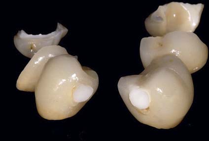
Fig 4-11 Veneered restoration with zirconia framework fractured due to insufficient dimension of the connector area.
To reduce the risk of framework fractures the current concept suggests the use of monolithic zirconia for tooth-supported FDPs, with facial microveneering. With a monolithic restoration it is less difficult to fulfill the minimal requirements of connector surface area of 12 mm2. The main indication for metal-ceramic tooth-supported FDPs today are large multiunit FDPs on periodontally compromised teeth with increased mobility. For such cases, high flexure strength is required for the framework material, making metal-ceramic the material of choice. Moreover, metal-ceramic is also recommended for tooth-supported cantilever FDPs with a cantilever unit of more than one tooth.
The overall 5-year failure rate of RBFDPs lost due to framework or material fractures is reported to be 1.7% and, as for tooth-supported SCs and FDPs, this complication is also material dependent10. Fractures are rarely reported for RBFDPs made of metal-ceramic, zirconia, and reinforced glass-ceramic. RBFDPs made of metal-resin, glass-infiltrated ceramic, and fiber-reinforced composite are significantly more often lost due to material fractures10.
To reduce the risk of a retainer fracture, the current concept recommends zirconia as framework material for RBFDPs.
Stay updated, free dental videos. Join our Telegram channel
VIDEdental - Online dental courses


