Introduction
Our objective was to determine the perception of smile esthetics among orthodontists and laypeople with respect to asymmetries on the maxillary incisor edges in a frontal smile analysis.
Methods
Two frontal close-up smile photos of 2 women, 1 white and 1 Afro-Brazilian, were selected for this study. Both smiles displayed healthy maxillary anterior dentitions. The images were digitally altered to create tooth wear on the maxillary left central and lateral incisors in 0.5-mm increments. The final images were randomly assembled into a photo album that was given to 120 judges, 60 orthodontists and 60 laypersons. Each rater was asked to evaluate the attractiveness of the images with visual analog scales. The data collected were statistically analyzed with 1-way analysis of variance with the Tukey post-hoc test and the unpaired Student t test.
Results
The most attractive smiles in both types of smiles were those without asymmetries and the 0.5-mm wear in the lateral incisor. In general, tooth wear was considered unattractive by both groups of raters following a pattern: the more tooth wear, the more unattractive the smile; tooth wear in the central incisor was considered more unattractive than in the lateral incisor. For both group of raters, 0.5 mm of wear in the central incisor was considered unattractive, whereas the thresholds for lateral incisor discrepancies were 0.5 mm for orthodontists and 1.0 mm for laypersons.
Conclusions
The result of this study corroborates the clinical assumption that symmetry between the maxillary central incisors is a paramount goal for esthetic treatments.
To obtain optimal esthetic results, it is of paramount importance for clinicians to follow esthetic guidelines. For many years, these parameters were based only on authors’ opinions rather than on evidence-based literature. These guidelines could be biased, since the concept of beauty has great subjectivity and is strongly influenced by the opinions of others. The literature suggests that orthodontists and laypeople have different perceptions of smile esthetics when evaluating a variety of orofacial characteristics, and that orthodontists are more sensitive in detecting deviations from ideal than the general public.
To provide more objective guidelines regarding the perception of smile esthetics, numerous studies were performed by using digital-image manipulations. Thereby, some smile characteristics were better elucidated: smile arc, amount of gingival display, type of buccal corridors, presence of dental and gingival asymmetries, presence of a midline diastema, influence of midline and long axis deviations, and importance of maxillary incisor sizes and proportions.
According to the literature, an esthetic treatment plan must begin at the maxillary central incisor area ; thus, dental or gingival asymmetries must be carefully analyzed. The impact of gingival asymmetries on the perception of smile esthetics is well documented in the literature. When there is a gingival margin discrepancy between the central and lateral incisors, neither laypersons nor dental professionals considered a 2-mm discrepancy unesthetic. However, when the gingival margin discrepancy was between the central incisors, slight discrepancies were considered unattractive by orthodontists and laypersons. Although this confirms that symmetry between the maxillary central incisors is an important aspect, it also highlights a question: if small gingival asymmetries are not recognized by laypeople as unattractive, is it necessary to treat?
After we analyzed this information, other questions arose. If an asymmetry is related to the maxillary incisor edges, what do laypeople and orthodontists perceive? In other words, what is the threshold for these people when evaluating uneven central and lateral incisors with tooth wear? This information is of paramount importance because it can assist the orthodontic clinician during the finishing and detailing phases. For instance, Kokich et al stated that if a dental asymmetry is not recognized as unesthetic, it might not be necessary to commit the patient to restorations.
Those questions were explored in this study, with the objective to determine the perception of smile esthetics among orthodontists and laypeople with respect to asymmetries on the maxillary incisor edges in a frontal smile analysis. The null hypothesis tested was that these asymmetries would be equally rated as attractive by orthodontists and laypeople.
Material and methods
According to a pilot study, a sample size calculation was undertaken by using software (version 5.0; BioStat, Mamirauá Institute, Tefé, Amazonas, Brazil). On the basis of a significance level of alpha 0.01 and the effect size estimated at 0.95, the sample size was calculated to achieve 80% power. This calculation showed that 58 subjects in each group were necessary.
Two standardized frontal photos of pleasant smiles of a white woman and an Afro-Brazilian woman were selected for this study. Both women had unworn, unrestored, and healthy maxillary incisors and had not had orthodontic treatment. These smiles were considered highly attractive and followed some principles of an ideal smile described in the literature: adequate width-to-length proportion of the esthetic zone, convex smile arc, gingival display less than 1.0 mm, gingival line of the central incisor matching the canine and the lateral incisor slightly below, and progressive increases in the depth of tooth embrasures from the central incisor to the canine.
The selected images were digitally altered by using Adobe Photoshop (CS3; Adobe Systems, San Jose, Calif). The photos were manipulated to produce symmetrical images and were then retouched to adjust color, brightness, and contrast, as well as to remove any discolorations on the lips and skin. Each image was then condensed to achieve an image with measurements identical to those on the actual patient. Thus, each millimeter measured on the digital and printed image was equivalent to each millimeter measured clinically on the patient, with the maxillary central incisor as the reference. Furthermore, after recommendations from previous studies, much of the nose and chin was removed to reduce the number of variables in the images.
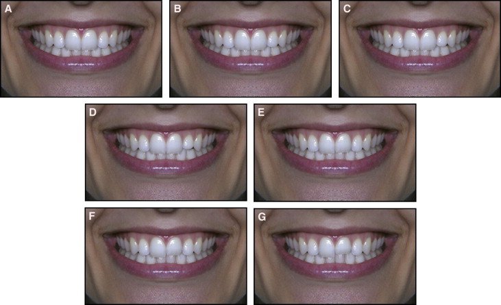
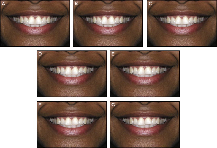
Each new image was altered in 0.5-mm increments on the incisor edges of the maxillary left central and lateral incisors. In all images, the gingival margins, the papillary heights, and the right sides of the image were not altered. For both photos, 7 new images were created ( Figs 1 and 2 ).
The final images were digital files with a resolution of 300 dpi. They were professionally printed with specialized digital equipment (Minilab Digital Frontier 570; Fuji Film, Manaus, Amazonas, Brazil) on standard A4 size format (29.7 × 42 cm) Kodak Edge Generations paper (Kodak do Brasil, Manaus, Amazonas, Brazil). Then a photo album was assembled containing all images from each group in random order.
The album was given to 120 judges, 60 orthodontists (37 men, 23 women) and 60 laypeople (32 men, 28 women) with a college education but no dental background. Each rater was given brief information about the study and asked to evaluate the attractiveness of the images. Along with the album, each judge received a form with 100-mm visual analog scales printed for each image, as in previous studies. The scale ranged from “very unattractive” on the far left to “very attractive” at the far right. A line was also printed at the midpoint of each scale to provide a reference line for an average level of attractiveness. All judges marked a point along the scale according to their perceptions of smile esthetics. The scores were then measured in millimeters by the first author with an electronic digital caliper (Starrett, Suzhou, China).
To assess the reliability of the method, 6 raters from each group were randomly selected. They were asked to evaluate 1 page of the album on which there were 2 identical images. Correlation coefficients were used to compare the scores for those images to determine intrarater agreement. High levels of reliability were found, since all coefficients were greater than or equal to 0.76 for both groups of raters.
Statistical analysis
The data were statistically analyzed with SPSS software (version 16.0; SPSS, Chicago, Ill). Descriptive statistics were reported as means and standard deviations. Differences in the mean esthetic scores in the levels of asymmetries were analyzed by using 1-way analysis of variance with the Tukey post-hoc test. To compare the distributions of the mean scores between orthodontists and laypersons, the unpaired Student t test was used. The level of signiflcance was established at 5%.
Results
From the orthodontists’ standpoint, the most attractive smiles among the images of the white woman were the symmetrical smile (mean, 86.0) and the 0.5-mm asymmetry in the lateral incisor (mean, 78.20); the least attractive was the 1.5-mm wear at the central incisor (mean, 9.27). From the laypersons’ opinions, the most attractive smiles were the symmetrical smile (mean, 87.26), the 0.5-mm wear on the lateral incisor (mean, 87.15), and the 1.0-mm wear on the lateral incisor (mean, 80.43); the least attractive smiles were those with 1.0 mm and 1.5 mm of wear on the central incisors (means, 29.34 and 22.36, respectively) ( Table I ).
| White woman | |||||||
|---|---|---|---|---|---|---|---|
| Altered asymmetry | Orthodontists | Laypersons | Difference | ||||
| Mean | SD | Results ∗ | Mean | SD | Results ∗ | ||
| No asymmetry | 86.00 | 9.99 | A | 87.26 | 9.71 | A | |
| 0.5 mm, lateral incisor | 78.20 | 8.45 | A | 87.15 | 8.31 | A | † |
| 1.0 mm, lateral incisor | 51.64 | 16.2 | B | 80.43 | 13.89 | A, B | † |
| 1.5 mm, lateral incisor | 37.04 | 16.05 | C | 61.91 | 10.92 | C | † |
| 0.5 mm, central incisor | 51.49 | 13.33 | B | 72.44 | 9.25 | B | † |
| 1.0 mm, central incisor | 22.35 | 10.82 | D | 29.34 | 14.29 | D | † |
| 1.5 mm, central incisor | 9.21 | 3.7 | E | 22.36 | 12.21 | D | † |
∗ Smiles with the same letter did not differ from each other ( P <0.05).
† statistical difference between the 2 group of raters ( P <0.05).
Similar results were found for the Afro-Brazilian woman’s smile. For the orthodontists, the most attractive smiles were the symmetrical smile (mean, 83.41) and the 0.5-mm wear on the lateral incisor (mean, 80.40), and the least attractive were those with 1.0 mm and 1.5 mm of wear on the central incisor (mean, 24.04 and 16.05, respectively). For the laypersons, the most attractive smiles were the symmetrical (mean, 87.61), the 0.5-mm wear on the lateral incisor (mean, 86.16), and the 1.0-mm wear on the lateral incisor (mean, 80.32); the least attractive were the smiles with wear of 1.0 mm and 1.5 mm on the central incisor (means, 29.18 and 23.98, respectively) ( Table II ).


