Introduction
Cone-beam computed tomography (CBCT) gives orthodontists and other dental clinicians 3-dimensional information for planning treatment in the craniofacial region. Often overlooked are incidental findings outside the treatment region of interest.
Methods
Two patients with incidental findings of skull-base abnormalities are presented. The orthodontic patient was tentatively diagnosed with a notochordal remnant in the clivus; the implant patient exhibited an empty sella turcica.
Results
For the clivus lesion in the orthodontic patient, an artifact was ruled out after a second CBCT image and further distinguished from a fat-containing tumor after magnetic resonance imaging. The impression after magnetic resonance imaging was a notochordal remnant, although chordoma was also included in the differential, warranting a 6-month follow-up magnetic resonance image to confirm the diagnosis. The CBCT study for the implant patient demonstrated an enlarged sella turcica. The impression after the magnetic resonance imaging was an enlarged and partially empty sella with no evidence of a pituitary mass.
Conclusions
Orthodontists and implant surgeons may come across incidental findings outside their area of expertise on CBCT scans, highlighting the importance of appropriate consultation with maxillofacial radiologists. Notochordal remnants may present as nonexpansile intraosseous low-density areas. The challenge in distinguishing these lesions radiographically with chordomas warrants follow-up to confirm a diagnosis. An empty sella is a noteworthy finding because of its potential for endocrine and neuro-ophthalmological disorders despite an asymptomatic presentation.
Cone-beam computed tomography (CBCT) is increasingly used by orthodontists and other dental practitioners. It provides volumetric information that is otherwise unavailable in standard radiographs. This aids in making accurate orthodontic diagnoses in 3 planes of space and provides localization of vital structures during other dental and surgical procedures. However, CBCT scans typically cover a field of view larger than the practitioner’s area of expertise. This leads to the possibility of overlooking incidental findings outside these regions of interest, even though the practitioner is responsible for evaluating the entire volume for pathology. Such findings may be significant and warrant further investigation. The literature has reported that about 25% of CBCT images taken for orthodontics and other dental purposes show incidental findings. Another study reports 701 “reportable” findings in 381 CBCT scans.
In medical radiology, the most common reason for malpractice litigation is missed lesions. Coupled with the frequency of incidental findings on CBCT volumes, this becomes an important consideration for the clinician. A study evaluating the efficacy of identification of maxillofacial lesions by orthodontists and orthodontic residents concluded with the recommendation that clinicians should involve a radiologist in the interpretation of CBCT volumes. It reported that training by an oral and maxillofacial radiologist significantly increased the clinicians’ detection rate of lesions, but even still, the 43% total of missed lesion errors is at odds with general radiology rates ranging from 4% to 13.5%.
The incidental findings may be skeletal or soft tissue, even though soft tissues are not ideally visualized because of insufficient contrast resolution. An in-depth knowledge of head and neck anatomy, as well as an awareness of the incidence of pathologic findings and false-positive findings in CBCT studies, is essential for clinicians using this technology.
This report of 2 patients demonstrates examples of abnormalities in the skull base. Patient 1 was an orthodontic patient who was tentatively diagnosed with a benign notochordal cell tumor (also called a notochordal remnant) in the clivus. Patient 2 was an implant patient who exhibited a partially empty and enlarged sella turcica.
Patient 1: benign notochordal cell tumor in the clivus
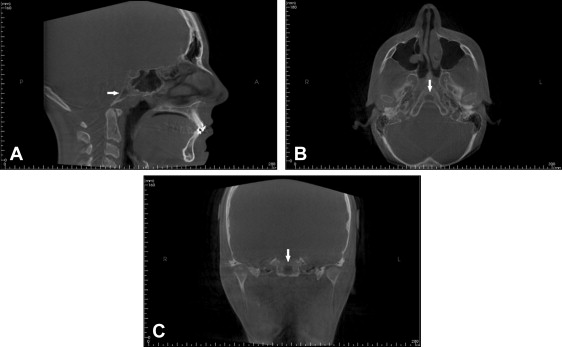
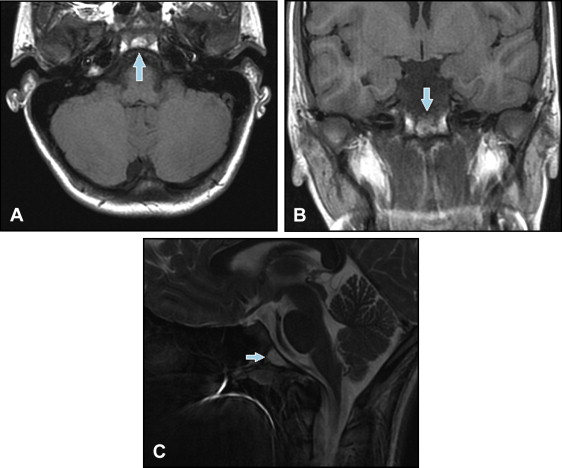
The patient, a 13-year-old girl, came to the orthodontist for evaluation. Her medical history was unremarkable. Her CBCT scan was referred to the Department of Oral and Maxillofacial Radiology at the University of Florida to overread for pathology and to rule out any need for medical or dental referral. A low-attenuation area in the clivus was noted. It was not possible to rule out an artifact from a lytic lesion. A follow-up CBCT image was ordered, and the possibility of an artifact was subsequently ruled out, since the low-attenuation area persisted ( Fig 1 ). A magnetic resonance imaging (MRI) examination and physician referral were advised. The MRIs showed an enhancing lesion in the body of the clivus ( Fig 2 ). The appearance suggested a notochordal remnant. Because of a potential similar presentation with a chordoma, a follow-up MRI in 6 months was advised to watch for any increase in size.


The follow-up CBCT and MRI images in this patient were indicated (1) for ruling out an artifact and (2) to monitor the lesion size. The significance of monitoring lesion size lies in the differentiation between notochordal remnants (also called benign notochordal cell tumors and ecchordosis physaliphora) and chordomas, which are locally invasive and destructive lesions. The vast majority of benign notochordal cell tumors remain intraosseous and exhibit mild osteosclerosis without bone destruction, whereas chordomas typically expand intraosseously and extraosseously with bone destruction. Furthermore, since benign notochordal cell tumors do not require surgical intervention unless they expand extraosseously, differentiation is important to avoid extensive surgical management and risk operating in the region of the clivus.
Patient 2: empty and enlarged sella turcica
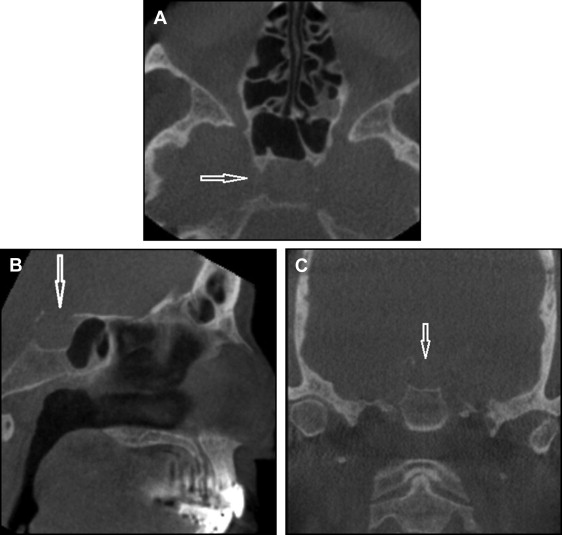
A 71-year-old woman came for implant evaluation. Her medical history was unremarkable. A preoperative CBCT was obtained and referred to the Department of Oral and Maxillofacial Radiology at the University of Florida for evaluation. The CBCT images showed enlargement of sella turcica with possible disruption of the posterior wall ( Fig 3 ). The MRI evaluation was advised to rule out a space-occupying lesion. The MRIs showed an enlarged sella turcica measuring 13 mm in its anteroposterior diameter. Sella was partially empty, with pituitary tissue flattened along the floor. No pituitary mass was seen.


