and Angela J. Yoon1
(1)
Columbia University College of Dental Medicine and Department of Pathology and Cell Biology, Columbia University Medical Center, New York, NY, USA
In this section we focus on focal and generalized gingival enlargements and gingivitis. When evaluating a gingival enlargement or nodule, it is prudent to take a radiograph of the area to ensure that the lesion is limited to the soft tissue.
|
Gingival lesions
|
|---|
|
Pyogenic granuloma
|
|
Peripheral ossifying fibroma
|
|
Fibroma/giant cell fibroma
|
|
Peripheral giant cell granuloma
|
|
Parulis
|
|
Eruption cyst
|
|
Gingival/alveolar cysts of the newborn
|
|
Localized juvenile spongiotic gingival hyperplasia
|
|
Mouth-breathing gingivitis
|
5.1 Pyogenic Granuloma
See Section 1.5 for entity details. Clinical photos of pyogenic granulomas of the gingiva are provided below.
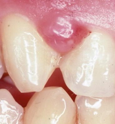
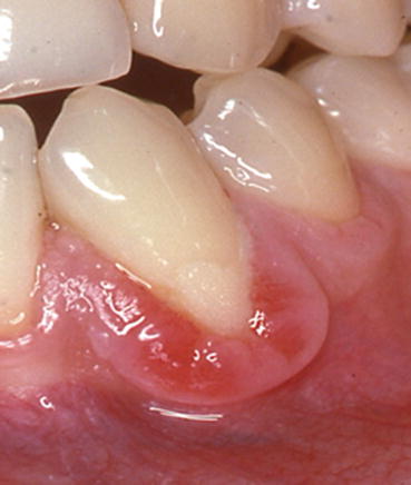
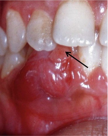
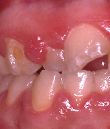
Fig. 5.1
Pyogenic granuloma. Erythematous exophytic nodule of the maxillary attached gingiva
Fig. 5.2
Pyogenic granuloma. Erythematous soft tissue growth attached to the mandibular gingiva
Fig. 5.3
Pyogenic granuloma. Erythematous and focally ulcerated (arrow) growth of the mandibular gingiva
Fig. 5.4
Pyogenic granuloma. Erythematous exophytic nodule of the maxillary attached gingiva
Fibroma
See page—for entity details. Photos of gingival fibromas are provided below.
There is a variant of fibroma referred to as a giant cell type fibroma. The clinical appearance is similar to a traditional fibroma except the surface can appear somewhat bosselated or papillary. It has a predilection for the gingiva and is not thought to be caused by chronic irritation or trauma. Treatment is surgical excision.
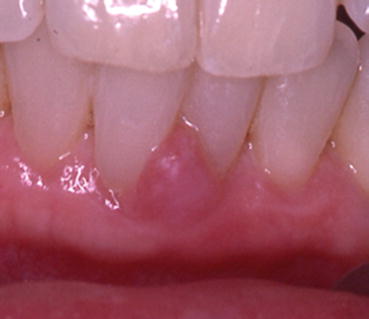
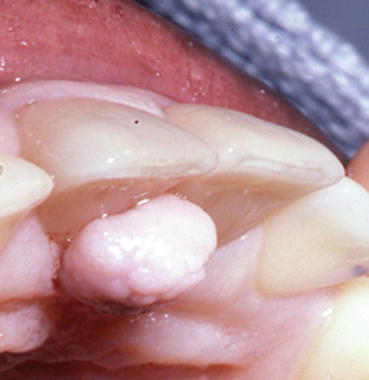
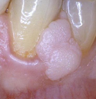
Fig. 5.5
Fibroma. Pink sessile nodule of the mandibular facial attached gingiva. The clinical differential diagnosis was fibroma vs. peripheral ossifying fibroma. (see Section 5.1)
Fig. 5.6
Fibroma, giant cell type. Pink sessile nodule of the maxillary lingual attached gingiva. Microscopic diagnosis was of the giant cell variant of fibroma
Fig. 5.7
Fibroma, giant cell type pink sessile nodule of the mandibular facial attached gingiva. Microscopic diagnosis was fibroma, giant cell type. Note the slight papillated appearance of the surface, which might cause the clinician to suspect a papilloma or condyloma
Clinical Note
The retrocuspid papilla is a circumscribed nodule resembling a fibroma located on the gingiva lingual to the mandibular cuspid. This entity is observed more frequently in young children and seems to regress with age. The retrocuspid papilla is considered to be a “normal anatomic structure” and should not be mistaken for a fibroma.
5.2 Peripheral Ossifying Fibroma
Clinical Appearance
Smooth, pink or red, exophytic nodule. Surface ulceration is not uncommon. Sessile or pedunculated. Nonpainful. Most commonly occurs in adolescents.
Etiology
Derived from cells of the periodontal ligament and thought to be a reactive lesion rather than a true neoplasm.
Location
Occurs on the gingiva only.
Differential Diagnosis
Fibroma, giant cell fibroma, if ulcerated and inflamed it could resemble pyogenic granuloma or peripheral giant cell granuloma.
Treatment
Surgical excision. In addition, any predisposing factors such as plaque or irritation should be removed. Recurrence is not likely but possible. Some authors have stated up to 20 % can recur.
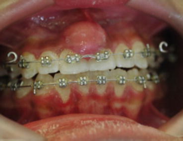
Fig. 5.8
Peripheral ossifying fibroma. Smooth surfaced, pink nodular mass of the maxillary anterior facial gingiva. (Photo courtesy of Dr. David Koslovsky, New York, NY)
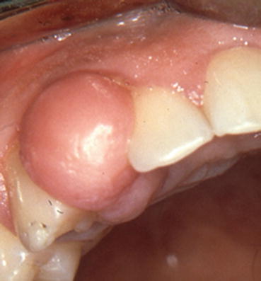
Fig. 5.9
Peripheral ossifying fibroma. Smooth surfaced, pink nodular mass of the maxillary anterior gingiva extending interproximally
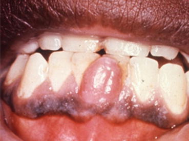
Fig. 5.10
Peripheral ossifying fibroma. Smooth surfaced, pink nodular mass of the mandibular anterior facial gingiva
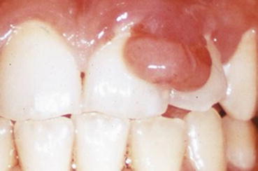
Fig. 5.11
Peripheral ossifying fibroma. Smooth surfaced, focally ulcerated pink-red nodular mass of the maxillary anterior facial gingiva. Clinically this lesion can be mistaken for a pyogenic granuloma
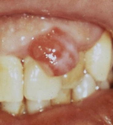
Fig. 5.12
Peripheral ossifying fibroma. Smooth surfaced, focally ulcerated pink-red nodular mass of the maxillary anterior facial gingiva
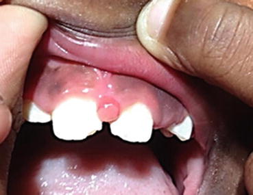
Fig. 5.13
Peripheral ossifying fibroma. Small smooth surfaced, centrally ulcerated pink-red nodule of the maxillary anterior facial gingiva
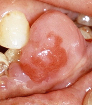
Fig. 5.14
Peripheral ossifying fibroma. Large smooth surfaced, pink-red nodular mass of the mandibular facial gingiva
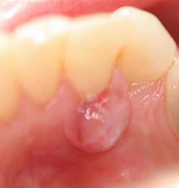
Fig. 5.15
Peripheral ossifying fibroma. Smooth surfaced, pink-red, focally ulcerated nodular mass of the mandibular lingual gingiva
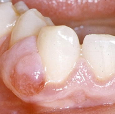
Fig. 5.16
Peripheral ossifying fibroma. Smooth surfaced, pink-red, focally ulcerated nodular mass of the mandibular lingual gingiva
Clinical Note
Peripheral ossifying fibroma can be clinically indistinguishable from a fibroma. If ulcerated and inflamed a peripheral ossifying fibromas can clinically mimic a pyogenic granulomas or peripheral giant cell granuloma.
Stay updated, free dental videos. Join our Telegram channel
VIDEdental - Online dental courses


