Introduction
Our aim was to investigate 8 years of vertical changes of the gingival margin and tooth displacement of maxillary molars and incisors in adolescents and adults.
Methods
Twenty-five adolescents and 10 adults were included in this study, with dental casts taken 2 and 10 years after orthodontic treatment. The gingival contour of the teeth was traced digitally using calibrated photographs of the 2 dental casts, and gingival changes were measured on crown superimposition. Eruption of the central incisors and first molar were measured on dental casts after scanning and superimposition on the palatal vault.
Results
Adults and adolescents presented a mean molar eruption of 0.27 and 0.34 mm and a mean incisor eruption of 0.39 and 0.73 mm, respectively. Adults and adolescents presented a mean molar gingival displacement of respectively 0.34 and 0.77 mm, and the adolescents a mean incisor gingival displacement of 0.44 mm. Correlation between secondary tooth eruption and gingival displacement was obtained only for the incisors in the adolescent group.
Conclusions
Adolescents and adults presented apical displacement of the gingival contour of the maxillary first molars, as was the case for maxillary incisors in adolescents. Secondary eruption of maxillary first molars and central incisors continues in adolescents and adults.
From childhood to adulthood, clinical crown heights in the maxilla increase differently according to the type of tooth. However, information about gingival displacement in healthy adults is lacking, particularly for posterior teeth, and we still do not know if gingival movements under healthy conditions stop during life.
Secondary tooth eruption (eruption after the teeth have reached the occlusal level) may play a role in the establishment of gingival position. In adults, the incisal edge of maxillary incisors is displaced in an occlusal direction by 0.95 mm in relation to the nasion over 20 years. Nevertheless, molar eruption has been measured rarely, and we do not know if and when molar eruption stops.
A relation between tooth eruption and gingival displacement has been shown for the maxillary central incisors, but no information exists regarding if this is the case for posterior teeth.
The aim of this retrospective longitudinal study was to monitor, on dental casts, 8 years of changes of the gingival contour of maxillary central incisors and first molars in adolescents and adults. We also measured secondary tooth eruption to determine if it is correlated to the gingival displacement.
The null hypothesis is: “no difference exists in gingival displacement and tooth eruption between maxillary central incisors and maxillary molars in either growing individuals or young adults.”
Material and methods
Subjects
Cases were selected among patients who finished orthodontic treatment at the Department of Orthodontics, University of Geneva, between 1984 and 1996 and who had dental casts taken approximately 2 and 10 years after the end of orthodontic treatment. Exclusion criteria comprised restoration of the maxillary central incisors and maxillary first molars performed or replaced during the follow-up period, poor quality of dental casts, and periodontal problems noticed by the practitioners at or between the 2 documentations.
Forty-two patients presented the necessary documentation. Seven were excluded, 3 because of inadequate quality of dental casts and 4 because of restorations.
According to the age of the patients at the first documentation used in the study (2 years after debonding and the end of orthodontic treatment), 2 groups were defined. The adolescent group comprised 25 subjects (7 boys, 18 girls), with a mean age of 15.9 years (range: 14.1 to 17.8). The adult group comprised 10 subjects (6 men, 4 women), with a mean age of 31.2 years (range: 21.4 to 46.4).
Measurements of gingival margin and displacement
On the first and second dental casts, teeth 15 and 16, teeth 25 and 26, and teeth 11 and 21 were photographed together in a standardized way. The occlusal plane was horizontal, and the contact point between both pair of teeth was centered in the photographs. The distance between the camera and the models was always the same to keep a constant and easily calculated magnification. This magnification has been calculated by photographing a ruler placed on the dental cast holder.
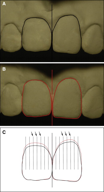
On the digital images, all teeth photographed were traced with drawing software (FreeHand 8.0, Macromedia Inc., San Francisco, Calif), and the line passing through the surface of contact of both teeth and perpendicular to the occlusal surface was also traced. Both drawings from each case were then superimposed, fitting the vertical placement of the drawing on the angles of the teeth. The measurements were done on the image with both drawings superimposed (Viewbox, version 3.1.1.12, dHAL Sofware, Kifissia, Greece) after magnification adjustment. The most mesial and the most distal points of each tooth were defined, and each tooth was divided into 8 equal parts, with lines parallel to the principal line. The intersections of these lines with the two tracings of the gingival contour were then recorded ( Fig 1 ). The gingival displacement on each line was calculated by the computer, and we calculated gingival displacement for each tooth by averaging the 3 gingival values in the center of the tooth.
Secondary eruption measurements
All the casts were scanned with the Laserscan 3D (Willytec, GmbH, Gräfelfing, Germany). The models were put on the articulated base with their occlusal plane parallel to the ground, and the image was then transferred to a computer (Siemens Expert, Siemens AG, Munich, Germany).
On the 3D model, we compared the position of the central incisors and the first molar at the first and second documentation. The structure of reference was the palatal vault, distal of the third rugae. All the points of reference were localized on easily reproducible areas of each tooth and were the same on both sets of casts.
Statistics
All the statistics were conducted using SPSS 13.0 for Windows (SPSS Inc., Chicago, Ill). Statistical significance was set to <0.05. The results of the adolescent and the adult group were compared by using an unpaired t test. The same test was used to compare measurements on incisors and molars. Pearson correlation coefficient was calculated to relate gingival displacement to secondary tooth eruption, molar-to incisor secondary eruption, and molar-to-incisor gingival displacement. Tooth and gingival displacement were evaluated using confidence intervals.
Error of the method
The error of the method for the gingival displacement was tested by measuring the photographs on 2 different occasions on 15 studied cases. The error of the measurements was calculated by using the Dahlberg’s formula ( Se2=∑d2/2n
Se 2 = ∑ d 2 / 2 n
) and ranged from 0.05 to 0.1 mm.
In order to compare our method with a method already described in the literature , the gingival displacement measured on the casts was compared with the 1 using intraoral photographs taken on the same day as the dental casts in 20 cases. The frontal intraoral photographs should have been taken symmetrically from a centric position parallel to the occlusal plane with the central incisors centered in the photograph. High correlation (Pearson correlation) was found between the 2 methods ( r = 0.802; P <0.01).
The method used for the measurements of tooth eruption was evaluated by double localization of the same points of reference with a difference of 10 weeks between the 2 measurements and calculated by Dahlberg’s formula. Standard error (SE) was found to be low (17.1 μm). The differences in the coordinates were assessed and compared using paired t test. A high repeatability of the localization of points of reference and a high correlation between the 2 sets of measurements were found that corresponds to a previous published study.
Stay updated, free dental videos. Join our Telegram channel
VIDEdental - Online dental courses


