Introduction
The placement and removal torques of mini-implants were evaluated as an index of implant stability. We examined factors affecting the initial and long-term stability of mini-implants.
Methods
We measured the placement and removal torques of 134 mini-implants placed in buccal posterior alveolar bone and assessed the relationships among placement and removal torques, placement period, age, sex, and cortical bone thickness. The mini-implants were machine-surfaced, 1.6 mm in diameter and 8 mm long. A torque screwdriver was used to measure the peak torque values.
Results and Conclusions
The placement and removal torques averaged approximately 8 and 4 N cm, respectively. A torque of 4 N cm suggests sufficient anchorage capability for mini-implants. No significant correlation between placement and removal torques was found. Placement torque was significantly related to age and cortical bone thickness in the maxilla, whereas removal torque was not significantly related to placement period, age, sex, or cortical bone thickness.
In recent years, orthodontic mini-implants have been used as orthodontic anchors. Orthodontic treatment has been advanced by the use of mini-implants; however, mini-implants are occasionally removed because they become mobile during treatment.
Many researchers have investigated the risk factors for failure of mini-implants to improve the success rate. They found that the primary stability of mini-implants is related to the mechanical characteristics of the interface between the mini-implant and bone in relation to factors such as bone quality and quantity, and screw diameter, length, and design.
To evaluate the initial stability of mini-implants, previous studies measured implant placement torque when tightening mini-implants. Those authors found that the recommended placement torque was 5 to 10 N cm for successful implantation, although they did not examine factors affecting the initial stability in detail. The removal torque reflects the characteristics of the implant-bone interface during and after long-term orthodontic treatment, and can also be used to evaluate the anchorage capability of mini-implants because removal torque is the resistance force required to remove a mini-implant after orthodontic treatment. Some researchers measured the removal torque of mini-implants in animals or bone specimens. Kim et al measured the removal torque of surface-treated mini-implants in humans.
In this study, we determined the placement and removal torques of machine-surfaced mini-implants in patients in relation to placement period, age, sex, and cortical bone thickness to identify factors that affect initial and long-term stability of mini-implants.
Material and methods
Fifty-seven orthodontic patients (148 implants) were studied. Fourteen implants that developed mobility and loosening during orthodontic treatment were excluded from the study. The final study group comprised 52 patients (10 male, with 25 implants; 42 female, with 109 implants), whose ages ranged from 13.9 to 63.5 years (average, 26.1 ± 8.4 years [mean ± SD]). Titanium mini-implants were placed in the buccal posterior alveolar bone in all subjects as anchors for orthodontic treatment at Nihon University Dental Hospital.
Computerized tomography (3D Accuitomo, J. Morita, Kyoto, Japan), with 0.125-mm slices with a voxel size of 0.125 mm in super-high-resolution mode, was used for diagnostic imaging of the area around the site before implant placement. The cortical bone thickness was measured at the prepared sites in the maxilla and mandible. The site of implant placement was identified by measuring the height from the archwire to the position of the implant on the tomogram at the prepared site in the interroot gap between the second premolar and the first molar, or the first molar and the second molar.
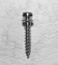
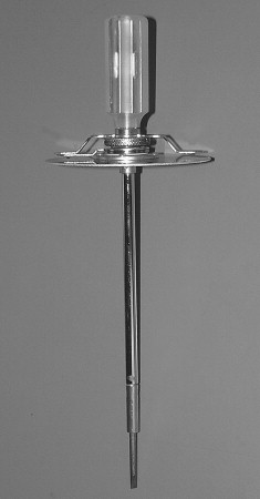
Titanium mini-implants measuring 1.6 mm in diameter and 8 mm in length (ISA orthodontic implants, Biodent, Tokyo, Japan) were used ( Fig 1 ). After local anesthesia was administered, a pilot hole was drilled with a bone drill into the buccal alveolar bone posterior to the second premolar or the second molar of the maxilla or mandible without raising a flap. To improve the success rate, we used bone drills with diameters of 1.0 mm in the maxilla and 1.3 or 1.4 mm in the mandible to control the placement torque, based on published results. For the mandible, a 1.3-mm or 1.4-mm diameter bone drill was chosen depending on bone stiffness; when the placement torque exceeded 10 N cm during placement of a mini-implant in a pilot hole 1.3 mm in diameter, the pilot hole was enlarged with the 1.4-mm bone drill. The peak value of the placement torque when tightening the mini-implant was measured by using a torque screwdriver (N2DPSK, Nakamura, Tokyo, Japan) ( Fig 2 ). The torque screwdriver has a round dial gauge with a pointer indicating the peak value, and the manufacturer stated that its accuracy is ± 3%. The peak placement torque was recorded at the final turn when the mini-implant was tightened into the implant hole.
Immediately after placement, an orthodontic force of less than 2 N was applied to the mini-implant. Each patient was prescribed an antibiotic for 3 days after placement to control infection. After clinical use of the mini-implant, the peak removal torque was measured by using the torque screwdriver when the mini-implant was removed. To verify the hypotheses that removal torque increases with greater placement torque, longer placement period, and thicker cortical bone, we investigated the relationships among the placement and removal torques, placement period, cortical bone thickness, sex, and age.
The Pearson correlation coefficient was calculated to evaluate the relationships among placement torque, removal torque, placement period, age, and cortical bone thickness. The paired t test was used to examine the difference between placement and removal torques. The unpaired t test was used to compare torque with sex and age. These analyses were performed with the SPSS statistical program (SPSS Japan, Tokyo, Japan); P <0.05 was considered significant.
This study was approved by the ethics committee of Nihon University School of Dentistry, and all patients consented to participate in this study.
Results
The rate of mobility and loosening of the mini-implants was less than 9.5%. There were no significant differences in the placement and removal torques between the right and left sides, or in cortical bone thickness. No significant correlation between the placement and removal torques was found in the maxilla (r = −0.063, P = 0.611) or the mandible (r = −0.083, P = 0.503). The removal torque was significantly lower than the placement torque in both the maxilla and the mandible ( Table I ). When the subjects were divided into 3 groups according to placement torque (low, 0-5; intermediate, 5-10; and high, 10-15 N cm) the removal torque did not change in the low-torque group, but it decreased significantly in the intermediate- and high-torque groups ( Table II ). No sex differences in the torques were found ( Table III ). Significant correlations were found between age and placement period, age and placement torque, and placement period and placement torque, whereas no parameters were correlated with the removal torque ( Table IV ). Placement torque was significantly related to maxillary cortical bone thickness, but removal torque was not related to cortical bone thickness ( Table V ). No significant correlation was found between torque and cortical bone thickness in the mandible ( Table VI ).
| Placement torque | Removal torque | Difference | |||||
|---|---|---|---|---|---|---|---|
| Mean | SD | Mean | SD | Mean | SD | n | |
| Female | 8.09 | 2.68 | 4.07 | 2.00 | −4.02 | 3.50 | 109 |
| Male | 7.80 | 2.16 | 4.93 | 2.17 | −2.87 | 2.97 | 25 |
| Placement period | Placement torque | Removal torque | Difference | |
|---|---|---|---|---|
| Age (y) | −0.299 ∗ | −0.287 ∗ | −0.087 | 0.165 |
| Mean, 26.1 | ||||
| SD, 8.4 | ||||
| Placement period (mo) | – | 0.195 ∗ | 0.101 | −0.086 |
| Mean, 23.1 | ||||
| SD, 6.7 |
Stay updated, free dental videos. Join our Telegram channel
VIDEdental - Online dental courses


