Introduction
The aims of this study were to investigate the effects of 2 force levels on the amount of relapse and to determine whether there is a relationship between the rates of tooth movement and relapse.
Methods
Approximately 20-g (group I) and 60-g (group II) forces were applied to the maxillary central incisors of 25 young adult (14 weeks of age) New Zealand female rabbits. Active tooth movement lasted 20 days. Then, the appliances were removed, and the incisors were released. The distance between the incisors was measured daily from the midlevels of the crowns by using a digital caliper during the active phase of tooth movement for 20 days, and then relapse was measured at the same level for 37 days. Analysis of variance and the Bonferroni multiple range test were used for statistical analyses.
Results
After active tooth movement, the mean total opening amounts were 3.98 ± 0.59 mm in group I and 4.82 ± 0.82 mm in group II, and the mean difference was approximately 0.8 mm. A rapid relapse was observed on the initial days in both groups, and its rate decreased with time. Significant relapse was observed in the first 5 and 8 days of the experiment in 20-g and 60-g force groups, respectively. The relapse in group II was significantly greater than in group I only on the first day of experiment. Statistically significant correlations were found between total tooth movement and relapse (R = 0.896, P <0.001).
Conclusions
These results showed a close relationship between the amount of relapse and orthodontic force magnitude. Greater relapse occurred during the initial days after appliance removal, and this indicates that retention appliances are needed immediately after the removal of orthodontic appliances.
Dental relapse, an undesirable outcome of orthodontic treatment, can be defined as a general tendency of the teeth to return their original positions after tooth movement. Relapse has been recognized as a major clinical problem among orthodontists, perhaps due to lack of understanding of the process, the mechanism of posttreatment relapse, and inadequate data regarding the effects of different treatment approaches on relapse tendency. The origin of this tendency is largely unknown or at least poorly understood, but it is generally accepted that intrinsic factors such as the periodontal ligament (PDL) and alveolar bone, and extrinsic factors such as growth of facial structures, soft tissue pressures, and interdigitation, might be potential factors in relapse.
Reitan evaluated the time required for recovery of different fibers after displacement in various areas of tooth roots of young dogs. He reported that some gingival fiber bundles remain displaced and stretched even after a retention period of 232 days. Periods of 83 and 147 days were required for rearrangement in the ligaments in the apical and middle regions, respectively. This study suggested that periodontal tissues have residual relapse potential even in a long term.
In orthopedic treatment, it has been shown that low force applications or slow displacements during orthopedic maxillary expansion can reduce the relapse potential. King and Keeling and King et al evaluated relapse and alveolar bone turnover in rats after removal of the orthodontic appliances. These studies showed that orthodontic relapse and bone remodeling continued for several days after removal of appliances. In an experimental study, van Leeuwen et al evaluated the effects of different intermittent forces on relapse and found no relationship between force magnitude and amount of relapse after active orthodontic tooth movement.
In clinical orthodontics, we usually use continuous forces for tooth movements. Nonetheless, the relationships between continuous orthodontic forces and amount of relapse have not been investigated sufficiently in the literature.
In the previous part of this study, the effects of force magnitude on tooth movement were discussed. Relationships between different continuous forces and amounts of relapse of orthodontically moved teeth are still unclear, and this question needs to be investigated. Therefore, the aims of this study were to examine the effects of 2 orthodontic forces on the amount of relapse and the relationships between the rates of tooth movement and relapse.
Material and methods
In the previous part of this study, the tooth movement period and the experimental design were explained. The study protocol was approved by the ethical committee board of the School of Dentistry, Atatürk University (protocol number is 2006/13), Erzurum, Turkey. Twenty-five young, healthy female New Zealand rabbits (mean age, 14 weeks) were used. The rabbits were randomly divided into 2 experimental groups, with 12 rabbits in group I and 13 rabbits in group II. The rabbits were individually housed in smooth-walled cages, and fed ad libitum with commercial pellets and water from thick-walled glass dishes. The mean weights of the animals were 2.72 ± 0.60 kg in group I and 2.97 ± 0.38 kg in group II at the beginning of the experiment.
The animals in each group were anesthetized at the first session by an intramuscular injection of ketamine (37.5 mg/kg) and xylazine (5 mg/kg). A small notch was made with a bur on the labial surface of the maxillary first incisors at 1.5 to 2 mm above the gingival margin, and then the notches were drilled in a vestibulo-palatal direction with a bur. Cooling was achieved with a syringe filled with physiologic saline solution.
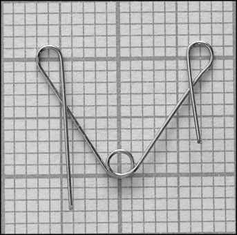
The appliance used in this study was an expansion spring. This spring was previously used by Storey and Stark and Sinclair, and modified by Karadede. The spring arms were 13 mm long with an angle of 70° ( Fig 1 ). To produce 2 forces, 0.012-in and 0.014-in round stainless steel archwires were used. The forces generated by the springs were measured with a gauge (040-713; Dentaurum, Ispringen, Germany) before application. When the free ends of the springs were closed to 4 mm, which corresponded to the width between the holes prepared in the rabbit incisors, the springs of the thin archwire initially exerted a force of 20 ± 3 g, and the other springs exerted a force of 60 ± 5 g. Springs exerting a force of 20 g were used in group I, and those with a 60-g force in group II.
The free ends of the springs were placed into the holes in the incisors. The residual ends were bent distally and cut to stabilize the springs in the mouth ( Fig 2 ).
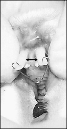
Active tooth movement lasted 20 days. Occlusal radiographs were taken from 2 rabbits in each group to observe whether sutural opening occurred. The springs were removed at the end of the 20th day. The incisors of all animals were allowed to relapse without any retention appliance. The distance between the incisors was measured every morning at the same time from the visible midlevel of the crowns ( arrows in Fig 2 ) by using a digital caliper with accuracy of 0.01 mm by the same author (M.E.).
To obtain reliable measurements, 3 successive measurements were made at each session, and their mean values were used for statistical analysis. Relapse measurements were made on all successive days until day 12. Because little relapse movement occurred during the remaining days, 3 measurements were made arbitrarily on days 16, 23, and 37.
Statistical analysis
To compare the amounts of relapse movement within and between groups, analysis of variance for repeated measurements was used. In addition, the changes in daily relapse movements were analyzed by the Bonferroni multiple range test.
All statistical analyses were performed with the Statistical Package for Social Sciences (Windows 98, version 10.0; SPSS, Chicago, Ill).
Results
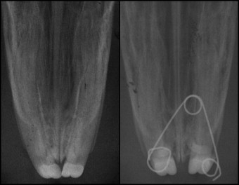
Tipping movements were observed in both groups at the end of active tooth movement, and there was no sutural opening in the animals of either group for whom occlusal radiographs were obtained ( Fig 3 ).
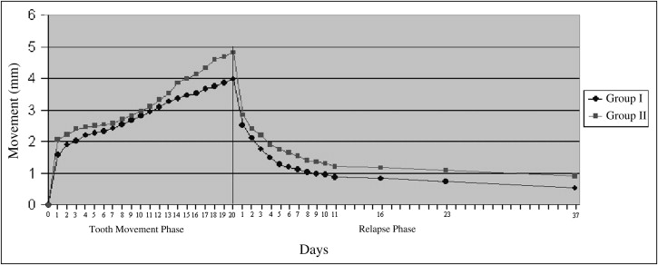
Daily measurements of the distances between the incisors are shown in Table I . The mean total amounts of opening were 3.98 ± 0.59 mm in group I and 4.82 ± 0.82 mm in group II, and the difference between the groups was approximately 0.8 mm at the end of active tooth movement. The mean total amounts of relapse were 3.45 ± 0.58 mm in group I and 3.89 ± 0.77 mm in group II. Time-displacement curves of tooth and relapse movements in both groups are shown in Figure 4 .
| Group I (20-g force, n = 12) | Group II (60-g force, n = 13) | |||
|---|---|---|---|---|
| Mean | SD | Mean | SD | |
| Day 0 | 3.98 | 0.59 | 4.82 | 0.82 |
| Day 1 | 2.52 | 0.48 | 2.85 | 0.64 |
| Day 2 | 2.12 | 0.36 | 2.41 | 0.57 |
| Day 3 | 1.82 | 0.31 | 2.15 | 0.57 |
| Day 4 | 1.52 | 0.21 | 1.90 | 0.46 |
| Day 5 | 1.33 | 0.18 | 1.74 | 0.45 |
| Day 6 | 1.20 | 0.17 | 1.61 | 0.43 |
| Day 7 | 1.12 | 0.17 | 1.54 | 0.42 |
| Day 8 | 1.03 | 0.16 | 1.41 | 0.38 |
| Day 9 | 0.99 | 0.17 | 1.36 | 0.37 |
| Day 10 | 0.96 | 0.18 | 1.31 | 0.35 |
| Day 11 | 0.88 | 0.17 | 1.21 | 0.33 |
| Day 16 | 0.83 | 0.22 | 1.18 | 0.31 |
| Day 23 | 0.73 | 0.18 | 1.09 | 0.28 |
| Day 37 | 0.53 | 0.30 | 0.92 | 0.32 |
| Total relapse | 3.45 | 0.58 | 3.89 | 0.77 |
The means and standard deviations of daily decreases in the gap between the incisors, and their within-group and between-group comparisons are shown in Table II . Rapid immediate relapses (37% in group I, 41% in group II) were observed on the first day after spring removal, and the rates gradually decreased during the following days. Relapse movements were statistically significant only during the first 5 days in group I and the first 8 days in group II ( Table II ).
| Days | Group I (20-g force, n = 12) | Group II (60-g force, n = 13) | Between-group significance |
||||
|---|---|---|---|---|---|---|---|
| Mean decrease | SD | Within-group significance |
Mean decrease | SD | Within-group significance |
||
| 1 | 1.46 | 0.24 | ‡ | 1.97 | 0.58 | ‡ | ∗ |
| 2 | 0.40 | 0.22 | † | 0.44 | 0.21 | † | NS |
| 3 | 0.30 | 0.11 | ‡ | 0.26 | 0.18 | ∗ | NS |
| 4 | 0.29 | 0.19 | ∗ | 0.25 | 0.19 | ∗ | NS |
| 5 | 0.19 | 0.09 | † | 0.17 | 0.11 | ∗ | NS |
| 6 | 0.13 | 0.13 | NS | 0.13 | 0.10 | ∗ | NS |
| 7 | 0.08 | 0.06 | NS | 0.11 | 0.08 | ∗ | NS |
| 8 | 0.09 | 0.07 | NS | 0.13 | 0.10 | ∗ | NS |
| 9 | 0.04 | 0.03 | NS | 0.05 | 0.03 | NS | NS |
| 10 | 0.04 | 0.03 | NS | 0.05 | 0.04 | NS | NS |
| 11 | 0.08 | 0.06 | NS | 0.10 | 0.06 | NS | NS |
| 16 | 0.05 | 0.12 | NS | 0.04 | 0.06 | NS | NS |
| 23 | 0.09 | 0.07 | NS | 0.08 | 0.08 | NS | NS |
| 37 | 0.21 | 0.25 | NS | 0.17 | 0.15 | NS | NS |
Stay updated, free dental videos. Join our Telegram channel
VIDEdental - Online dental courses


