Introduction
In this study, we examined the effect of neonatal administration of capsaicin on the magnitude of orthodontic tooth movement in rats.
Methods
Twelve timed pregnant Sprague-Dawley rats were randomized between the capsaicin group and the vehicle group. The pups received treatment with either capsaicin or vehicle on day 2 of life. Capsaicin treatment has been shown to produce a selective destruction of fine myelinated and unmyelinated Aδ and C sensory nerve fibers, causing an inhibition of the effects from neurogenic inflammation. Tooth-movement experiments began at 12 weeks of age. A mesial tipping force was applied to the maxillary first molar by using a 3-mm length of Sentalloy closed-coil spring (Dentsply GAC Intl, Bohemia, NY) activated from a bonded molar cleat to the maxillary incisors; this appliance delivers a constant tipping force of 50 g. Diastema measurements between the first and second molars were made at 2 and 4 weeks after appliance placement. Measurements were made indirectly from stone models by using a charge-coupled device microscope camera and Optimas 5.2 measurement software (Media Cybernetics, Bethesda, Md). Two-way repeated-measures analysis of variance (ANOVA) was used to analyze the differences between the groups.
Results
The capsaicin-treated rats and the controls did not differ in the amount of tooth movement at the collected time points ( P >0.05). Similarly, the magnitude of change of tooth movement from 2 to 4 weeks did not differ between the groups ( P >0.05). An increase in average diastema size was observed between 2 and 4 weeks after appliance activation in both treatment groups ( P <0.0001).
Conclusions
These results suggest that neonatal capsaicin desensitization in the rat does not affect the rate of orthodontic tooth movement after the application of a 50-g tipping force to the maxillary first molar. This might be due in part to the development of compensatory mechanisms in the chronically desensitized rat. Further studies are necessary to determine the reproducibility and histologic characteristics of this treatment.
Recent experimental studies have suggested that peripheral nerve fibers play a regulatory role in the control and development of local inflammatory reactions observed during orthodontic tooth movement (OTM). Calcitonin gene related peptide (CGRP) and substance P are two known multi-potent neuropeptides that contribute to the neurogenic component of inflammation by having multiple vascular, inflammatory, and cellular effects. An increased density of CGRP- and substance P-immunopositive nerve fibers has been observed in the pulp and periodontal tissues of mechanically stressed teeth. This coincides with the vascular and cellular changes observed during OTM. Conversely, the deprivation of sensory nerve supply has been shown to attenuate the local inflammatory responses after the application of an orthodontic force. Miller et al evaluated the effect of surgical transection of the maxillary nerve on OTM. Although no significant differences were found in the amount of tooth movement after the application of an orthodontic force, the denervated group had a smaller increase in the magnitude of tooth movement compared with the controls. Duan et al showed that denervation of dental tissues reduces the amount of bone formation. Collectively, these studies have suggested that neurogenic mechanisms might have a significant effect in the cellular processes that mediate tooth movement.
Chemical denervation with capsaicin treatment is a suitable method for analyzing the efferent actions of sensory nerves. Capsaicin has been extensively used as a tool in sensory neuron biology. Its neurotoxic actions, when applied systemically in high doses to neonatal rats, have been well documented. Studies by Jancsó et al have demonstrated that the administration of capsaicin to newborn rats results in the selective degeneration of B-type primary sensory dorsal root ganglion cells, giving rise to unmyelinated (Aδ and C) and myelinated fibers of small diameter. This produces depletion of neuropeptides from the central and peripheral parts of primary sensory neurons, causing inhibition of the effects from neurogenic inflammation. The capsaicin receptor, transient receptor potential vanilloid 1 (TRPV1), is a well-characterized cation channel expressed predominantly in unmyelinated sensory C fibers and activated by various noxious stimuli. TRPV1 is activated not only by capsaicin but also by protons or heat (with a threshold of approximately >43°C), both of which cause pain in vivo. Systemic administration of capsaicin to neonatal mice has been documented as a well-established method to ablate TRPV1-expressing sensory neurons. More recent studies have shown a relationship between TRPV1 receptors and inflammatory mediators released from the cyclooxygenase pathway, such as prostaglandin E 2 (PGE 2 ) and prostaglandin I 2 (PGI 2 ). Moriyama et al found that, in the presence of PGE 2 and PGI 2 , the temperature threshold for TRPV1 activation was reduced below 35°C, so that temperatures near body temperature were sufficient to activate TRPV1. In addition, an increase in the potentiation or sensitization of TRPV1 activity was observed through PGE 2 and PGI 2 receptor activation, leading to thermal hyperalgesia and inflammatory nociceptive responses. These studies further document the potential role of vanilloid capsaicin receptors in the inflammatory response.
The purpose of this study was to evaluate the effect of capsaicin-induced sensory denervation on the rate of OTM in Sprague-Dawley rats. These results would be of clinical importance, especially in patients when combined orthodontic and extensive surgical treatment is planned and sensory innervation is severed. Sensory nerve damage and paresthesia have been common findings in patients undergoing Lefort and mandibular ramus surgical procedures. If capsaicin-induced sensory denervation is shown to inhibit the rate of OTM, this would not only open a new avenue of research, but also suggest that special considerations should be taken in the orthodontic treatment of our orthognathic surgical patients who have been deprived of their sensory innervation.
In this study, we attempted to answer the question: “Will teeth under a constant orthodontic force move at the same rate if their sensory input is compromised by capsaicin desensitization at a neonatal age?”
Material and methods
The animals used in this study were 12-week-old (weight, 350 ± 25 g) male Sprague-Dawley rats raised and treated as described below in the animal facilities at the University of Minnesota. The animals were housed in accordance with National Institutes of Health guidelines and kept in a vivarium maintained at 22°C with a 12-hour alternating light and dark cycle. They were fed powdered rodent chow and water ad libitum throughout the study. All procedures were approved by the Animal Care and Use Committees at the University of Minnesota.
Twelve timed pregnant Sprague-Dawley rats were housed individually until delivery. Six pregnant rats were randomly assigned to the capsaicin group, and the remaining six rats were assigned to the vehicle group. One hundred fifty-six pups were obtained from the twelve pregnant rats. The average litter size was 13 pups. Six litters were assigned to the capsaicin group (75 pups), and the remaining six litters were assigned to the control group (81 pups). Average weight at day 2 after birth was 7.4 g.
Pups from the mothers assigned to the capsaicin group were injected with capsaicin (50 mg/kg, 5 μL/g; Sigma, St. Louis, Mo) on the second day of life, according to the protocol of Jancsó and Király. Capsaicin (1%) was dissolved in a mixture of 10% ethanol and 10% Tween 80 (Sigma Ultra) in 0.9% sodium chloride. The pups were injected subcutaneously in the dorsal thoracic region by pinching the skin on the back and inserting a 30-gauge needle on a 1-mL syringe. The pups in the control group received subcutaneous injections of the vehicle without capsaicin at day 2 after birth. The pups were returned to their respective mothers after treatment to ensure nurturing. The animals were weaned and sex differentiated at 28 days of age.
At weaning, 61 rats (81%) survived in the capsaicin group, and 77 rats (95%) survived in the control group. Only male rats were included in our study, since a previous study indicated that OTM varies depending on the phase of the estrous cycle in female rats. Specifically, cyclic changes in the estradiol level have been found to be associated with the estrous-cycle-dependent variations in tooth movement through its effects in bone resorption. A total of 73 male animals who survived the treatments were used in our study: 35 rats from the capsaicin group and 38 from the control group.
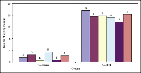
The animals’ responsiveness to noxious chemical stimuli was determined at 6 weeks of age by using the standard eye-wipe test, a method described by Szolcsány and Jancsó. A cotton-tipped applicator saturated with a solution of 0.01% (w/v) capsaicin was applied to the cornea in one eye, and the number of wiping motions that occurred in the subsequent 30 seconds was counted. This method has proved to be highly reliable in determining the effectiveness of capsaicin desensitization. In normal animals, this treatment induces a brief period of scratching or redness. The lack of a wiping response or casual wiping indicates desensitization.
General anesthesia for the placement of the orthodontic appliances was induced with the administration of 50 mg per kilogram of ketamine hydrochloride and 10 mg per kilogram of xylazine hydrochloride injected as one bolus dose intraperitoneally. If anesthesia was not effective within 15 minutes, a second injection of 150 μL of nembutal was given intraperitoneally. This gave adequate anesthesia for approximately 30 to 45 minutes. The rats were weighed before each anesthesia event to monitor their ability to thrive during this experiment.
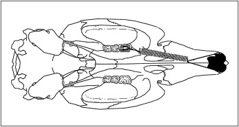
The rat model used in this study was modified from a model described initially by King et al. This model was used in a recent experimental study by Miller et al, with a detailed description of the method for the orthodontic appliance used in this study. A schematic representation of the orthodontic appliance is shown in Figure 1 .
Tooth movement measurements were taken at 2 and 4 weeks after appliance placement. The rats were anesthetized in a manner similar to that described by Miller et al. Impressions of the diastema distal to the maxillary first molar were taken with Vinyl Polysiloxane Imprint II wash material low viscosity and Garant mixing tips (both, 3M ESPE, St Paul, Minn) with intraoral injectable attachment tips. This material was allowed to set for 4 minutes before removing it from the mouth. Impressions were stored for 1 day in a temperate room before pouring with stone. According to the manufacturer’s specifications, no appreciable expansion or contraction will be observed with these storage times or conditions. The stone used to pour the impressions was Fujirock II (GC America Inc, Alsip, Ill). The stone was allowed to sit for 24 hours before separation from the impression material.
In preparation for the measurement procedures, the stone models were randomized and assigned code numbers. All measurements were done by one examiner (J.D.) who was blinded to the experimental conditions. A Navitar macro zoom 18-108 lens (Navitar Inc, Rochester, NY) attached to an MTI 3 charge-coupled device camera (DAGE-MTI Inc, Michigan City, Ind) was used to measure the diastemas. A laminated millimeter ruler was digitized at a constant focal length for calibration purposes. Diastemas were measured indirectly from acquired images of the stone models with the camera and Optimas 5.2 measurement software (Media Cybernetics, St. Louis, Mo). A measurement tool in the software program allowed us to calculate linear distances on the stone models from the calibrated ruler algorithm. Pilot experiments showed that this technique provided reproducible and accurate measurements.
Statistical analysis
Data from the eye-wipe test were expressed as mean ± standard error of the mean. The mean nociceptive thresholds were compared between groups by using a mixed-effects analysis of variance (ANOVA) in which the groups were a fixed effect, litters in the groups were a random effect, and rats in the litters were a second random effect. The means and standard errors of body weight were similarly calculated at baseline and on days 14 and 28. Differences in body weight between groups and time points were analyzed by using mixed-effects ANOVA with rats as the random effect. The diastema measurements were collected in triplicate from the stone models with the camera and the software program. The averages of the 3 measurements were calculated for each rat at both 14 and 28 days. Repeated measures ANOVA was used to analyze the differences between groups. The analysis was a mixed linear model with rats as the random effect and included these fixed effects: treatment group, time, and their interaction. The analyses were done with a statistical software package (version 8.2, SAS, Cary, NC) by using the mixed procedure and the restricted-likelihood method.
Results
Ophthalmic administration of capsaicin evoked a characteristic wiping response in the vehicle-treated rats at 6 weeks of age. Although ophthalmic instillation of capsaicin in the capsaicin-treated rats also evoked a wiping response, the number of wipes (1.8 wipes on average) was significantly less than in the corresponding vehicle-treated rats (15.7 wipes on average) of the same age ( P <0.0001) ( Fig 2 ).
Seventy-three animals started the tooth movement experiment at 12 weeks of age. Of these, 6 animals (2 capsaicin-treated and 4 vehicle-treated rats) had debonded appliances 2 weeks after their placement. These animals were excluded from the analysis. One additional animal from the capsaicin group had a debonded appliance on day 28; therefore, 28-day measurements were not taken for this animal. The major reason for bond failure was the debonding of the molar cleat from the occlusal surface of the molar at the enamel-resin interface. A total of 66 animals (90%) had intact appliances on day 28: 32 from the capsaicin group and 34 from the control group.
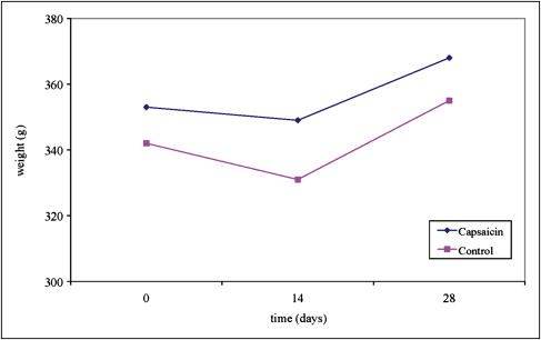
Sixty-six animals completed this study. Analysis of weight data indicated that the rats lost on average 7.8 g (2.2% decrease) in the first 2 weeks after appliance placement and gained 21.3 g in the subsequent 2 weeks. Averaging over the 2 groups, the differences in weight were significantly different among the 3 measurement times (time main effect, P <0.0001). Weight gain differences between the treatment groups were not significant (group main effect, P = 0.16). In addition, no significant difference in body weight change was found between the 2 treatment groups during the experiment (group-by-time interaction, P = 0.41) ( Fig 3 ). The initial reduction in body weight could be associated with the discomfort that the animals experienced as a result of initial appliance activation. However, a significant increase in body weight was observed at day 28; this was consistent with the biocompatibility of the appliance system and the ability of the animals to thrive.