Introduction
In growing patients with skeletal discrepancies, early diagnosis, evidence-based explanations of etiology, and assessment of functional factors can be vital for the restoration of normal craniofacial growth and the stability of the treatment results. The aims of our study were to compare the 3-dimensional pharyngeal airway volumes in healthy children with a retrognathic mandible and those with normal craniofacial growth, and to investigate possible significant relationships and correlations among the studied cephalometric variables and the airway morphology in these children.
Methods
Three-dimensional airway volume and cross-sectional areas of 27 healthy children (12 boys, 15 girls; mean age, 11 years) were measured by using cone-beam computed tomography volume scans, and 2-dimensional lateral cephalograms were created and analyzed. The subjects were divided into 2 groups based on their ANB angles (group I: 2° ≤ ANB ≤ 5°; group II: ANB >5°), and cephalometric variables, airway volumes, and cross-sectional measurements were compared.
Results
There were statistically significant differences in the following parameters: height of the posterior nasal plane ( P <0.05), pogonion to nasion perpendicular distance ( P <0.01), ANB angle ( P <0.01), mandibular body length ( P <0.01), facial convexity ( P <0.01), and total airway volume ( P <0.05). No statistically significant differences between the 2 groups were found in the cross-sectional area and the volumetric measurements of the various sections of the airway except for total airway volume, which had larger values in group I ( P <0.05).
Conclusions
The mean total airway volume, extending from the anterior nasal cavity and the nasopharynx to the epiglottis, in retrognathic patients was significantly smaller than that of patients with a normal anteroposterior skeletal relationship. On the other hand, differences in volume measurements of the 4 subregions of the airway were not statistically significant between the 2 groups.
Editor’s summary
When you examine a patient with a Class II malocclusion and a retrognathic mandible, do you assume that he or she has reduced airway capacity? If so, on what do you base that assumption? The aims of this retrospective, cross-sectional study were to compare pharyngeal airway volumes of healthy children who had retrognathic mandibles with those of children who had normal craniofacial relationships and to investigate the correlation between cephalometric variables and airway morphology in these children.
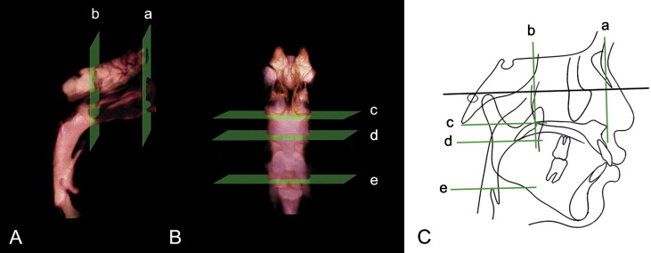
Pharyngeal airway structures were studied in 27 children (mean age, 11.19 years) who were referred for orthodontic treatment. Cone-beam computed tomography (CBCT) scans were obtained of all subjects for volumetric rendering for airway and cephalometric analysis. Two-dimensional (2D) cephalometric images were derived from the 3-dimensional (3D) CBCT scans, and the images were imported into a cephalometric software program for conventional 2D analysis. Volumetric renderings were acquired with InVivoDental software (Anatomage, San Jose, Calif).
Because this was a retrospective study, few subjects were available; therefore, this should be considered a pilot study. The results showed that healthy preadolescents with retruded mandibles have decreased total pharyngeal airway volumes. Future investigations of longitudinal airway changes in patients with different skeletal patterns and assessments of their craniofacial growth with 3D superimpositions will allow better understanding of the relationship between respiratory function and craniofacial morphology.