Introduction
The objective of this study was to investigate the in-vitro fluoride release from a glass ionomer orthodontic bonding system (Fuji I, GC Corporation, Tokyo, Japan) over a 2-month period and the in-vivo enamel fluoride uptake after 6 months.
Methods
Ten metal brackets were bonded with either glass ionomer or composite resin (Transbond XT, 3M Unitek, Monrovia, Calif; Light Cure), which served as controls, to recently extracted molars. The bonded teeth, cut at the level of their roots, were stored in distilled water that was renewed after every fluoride measurement at 1, 3, 7, 30, and 60 days. The in-vitro fluoride release was measured by using a fluoride ion-selective electrode, connected to an ion analyzer. Fifteen pairs of premolars were bonded with metal brackets with either the Fuji or the Transbond adhesive. Six months later, the teeth were extracted for orthodontic purposes, embedded in resin, and cross-sectioned, and the fluoride compositions between the outer and bulk enamel surfaces were evaluated with scanning electron microscopy and energy dispersive analysis. The results were analyzed with nonparametric 1-way analysis of variance (ANOVA) on ranks for in-vitro fluoride release and nonparametric 2-way ANOVA on ranks for in-vivo fluoride enamel uptake; group differences were investigated with the Holm-Sidak test at the .05 level. The Spearman rank correlation coefficient test was used to investigate the association between fluoride and aluminum levels in the interfaces of the specimens bonded.
Results
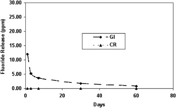
The initial burst of fluoride release observed for the Fuji adhesive after the first day of the experiment had a significant decrease with time, and it persisted throughout the monitoring period (60 days) ( P <0.05). Fluoride concentrations were found in both the outer and deeper enamel surfaces, with the outer sites having 4 times higher fluoride relative to the bulk for the glass ionomer ( P <0.05), and higher fluoride was found in the outer layers for the glass ionomer bonded enamel specimens ( P <0.05). However, the concurrent identification of aluminum and fluoride traces in the enamel implied that the source of this high fluoride concentration originated from cement particles and not from ionic uptake.
Conclusions
The short-term fluoride release and the absence of documented enamel uptake suggest that the glass ionomer orthodontic adhesive tested might provide protective action only through the reservoir mechanism.
Editor’s comment
The anticariogenic potential of glass ionomer cements has been shown to be effective in several in-vitro studies because of their ability to release fluoride. However, in-vivo evidence is limited regarding the amount of fluoride released from this material, and what has been published was primarily derived from short-term periods of monitoring. These authors investigated in vitro the duration of fluoride release from an orthodontic glass ionomer adhesive (Fuji I) and evaluated in vivo the depth of enamel fluoride uptake. The article is well written, and the authors properly analyzed and compared their findings with the results of other studies in the discussion. Their findings are based on the results of only 1 commercial type of glass ionomer adhesive (Fuji I), and therefore the conclusion might not represent all types of glass ionomer adhesives. The study would be even more useful if the authors had compared fluoride release and enamel uptake from glass ionomer adhesives of different manufacturers (eg, Ketac-Cem [ESPE] and Aqua Cem [Dentsply]) and from resin-modified glass ionomer adhesives. However, because of the recent scarcity of studies with a similar design and the high clinical interest of this topic, the following conclusions are a welcome addition to the literature.
The results confirm that the glass ionomer orthodontic adhesive releases fluoride in vitro when compared with a composite resin as the control, but this fluoride decreases progressively, leading to a short-term effect. The issue of fluoride release is closely related to its uptake from dental tissues. The results of the in-vivo portion of this study demonstrate that, after 6 months in the oral cavity, no fluoride was uptaken by the enamel, indicating that the beneficial action of fluoride might be due to the reservoir mechanism and not fluoride uptake.


