Introduction
Computer software can be used to predict orthognathic surgery outcomes. The aim of this study was to subjectively compare the soft-tissue surgical simulations of 2 software programs.
Methods
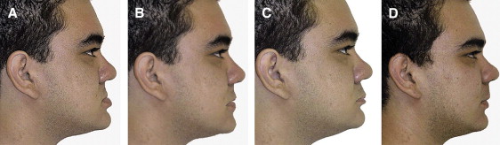
Standard profile pictures were taken of 10 patients with a Class III malocclusion and a concave facial profile who were scheduled for double-jaw orthognathic surgery. The patients had horizontal maxillary deficiency or horizontal mandibular excess. Two software programs (Dentofacial Planner Plus [Dentofacial Software, Toronto, Ontario, Canada] and Dolphin Imaging [version 9.0, Dolphin Imaging Software, Canoga Park, Calif]) were used to predict the postsurgical profiles. The predictive images were compared with the actual final photographs. One hundred one orthodontists, oral-maxillofacial surgeons, and general dentists evaluated the images and were asked whether they would use either software program to plan treatment for, or to educate, their patients.
Results
Statistical analyses showed differences between the groups when each point was judged. Dolphin Imaging software had better prediction of nasal tip, chin, and submandibular area. Dentofacial Planner Plus software was better in predicting nasolabial angle, and upper and lower lips. The total profile comparison showed no statistical difference between the softwares.
Conclusions
The 2 types of software are similar for obtaining 2-dimensional predictive profile images of patients with Class III malocclusion treated with orthognathic surgery.
Editor’s comment
Over the past 10 years, you have probably become accustomed to using 2-dimensional (2D) predictive imaging to help surgical patients evaluate expected facial changes. With recent improvements in some imaging programs, you’ve decided to upgrade to a popular program, either Dentofacial Planner Plus or Dolphin Imaging. However, these programs work differently, and it is possible that their predicted images will also differ. Your question is: “which program will provide the most accurate predictions?” If you are considering a change, you’ll want to read this article.
Ten orthognathic surgical patients who had mandibular retropositioning and maxillary advancement to correct a Class III malocclusion had photos taken before and after surgery. The predictions were based on pretreatment photographs but were taken 6 months after the surgery and used the actual jaw movements as the values for the predictions. A total of 101 orthodontists, oral and maxillofacial surgeons, and general dentists evaluated the images and rated the following items on a 5-point scale from similar to different: tip of the nose, nasolabial angle, upper lip, lower lip, menton region, base of the mandible, and complete profile.
The authors quickly determined that a precise evaluation of surgical simulation images is not easy, and there is a subjective aspect to these evaluations. Two-dimensional simulation of treatment effects has been our primary focus for many years. Variations in upper and lower lip morphology and the significant differences in response to both dental and skeletal changes appear to limit most software packages. This study seems to confirm what has been shown in many other studies, and the authors concluded that these softwares have similar limitations. The programs were not as good at predicting the outcome in Class III patients treated with bimaxillary osteotomy as they were for Class II patients. This could be because most Class II treatment requires only single-jaw osteotomy. Statistical results showed a certain similarity between the 2 programs with regard to evaluation of the profile prediction in 2 dimensions for Class III patients treated with bimaxillary surgery.