Introduction
The aim of this prospective in-vivo study was to investigate the possible effects of temperature changes from various adhesive cleanup procedures on pulpal tissue.
Methods
The materials, consisting of 40 sound maxillary and mandibular premolars to be extracted during orthodontic treatment, were randomly assigned to 4 groups, with 1 group as the control. The teeth in the 3 study groups were etched; brackets were bonded and then debonded. The remaining adhesive was removed with a tungsten carbide bur by using a high-speed hand piece. The teeth in the control group were not etched and bonded. In group 1, the residual adhesive was removed with water cooling, and the teeth were extracted 24 hours later. In group 2, the residual adhesive was removed without water cooling, and the teeth were extracted 24 hours later. In group 3, the residual adhesive was removed without water cooling, and the teeth were extracted 20 days later. The teeth were prepared for histologic examination, and the number of vessels, vessel areas and perimeters, extravasation of red blood cells, vascular congestion, and inflammatory cell infiltration were evaluated to determine pulpal tissue changes.
Results
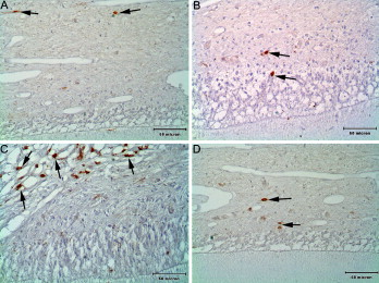
According to the findings from histologic and immunohistochemical evaluations, the coronal pulps of the teeth in groups 1 and 3 were almost similar to the control teeth, but some distinct pathologic changes were observed in group 2.
Conclusions
Adhesive removal without water cooling caused some vascular and pulpal tissue alterations, but these were tolerated by the pulpal tissues, so the changes were reversible.
Editor’s comment
The effect of resin grinding on hard dental tissue physiology has been investigated in the past. Likewise, a number of in-vitro studies have examined pulpal temperatures during this process. These Turkish authors performed the first in-vivo study of the effects of debonding-induced heat on the histology of pulp. Their subjects included patients whose treatment plans included extractions. Before the extractions, the authors bonded brackets to the teeth and then debonded them by 3 modes. In group 1, the residual adhesive was removed with water cooling, and the teeth were extracted after 24 hours. In group 2, the residual adhesive was removed without water cooling, and the teeth were extracted after 24 hours. In group 3, the residual adhesive was removed without water cooling, and the teeth were extracted after 20 days. It would have been useful to include 1 more group, in which water cooling was followed by a 20-day period; this would have provided additional evidence and completed the picture of the variables of cooling or delayed histologic changes. The histologic examination of the teeth included the number of vessels, vessel areas and perimeters, extravasation of red blood cells, vascular congestion, and inflammatory cell infiltration. The lack of quantitative data for some variables tested makes it difficult to extrapolate a clear-cut picture of the findings. However, the results primarily showed that the coronal pulps of the teeth in groups 1 and 3 were almost the same as the control teeth, but some distinct pathologic changes were observed in group 2. Adhesive removal without water cooling caused some vascular and pulpal tissue alterations, but these were tolerated by the pulpal tissue, and the changes were reversible. These findings establish the lack of significant hazardous effects of debonding on pulp vitality. However, because of the unpredictability of pulp response, care should taken when using techniques such as water cooling during resin grinding.


