Fig. 11.1
Color virtual model
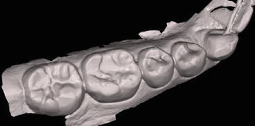
Fig. 11.2
Monochromatic virtual model
Open and closed systems.
-
Open – digital impression file (usually .stl file) can be imported into other software programs for design and restoration fabrication.
-
Closed – digital impression file stays within the same software system for design and restoration fabrication.
Most systems employ continuous image capture or true video during the scanning process.
Proven accuracy (Ahrberg 2016).
Secure virtual submission (Digital Dental Exchange 2016).
11.1.1 Capabilities of Intraoral Scanners
-
Magnified image and ability to zoom allow better visualization of tooth preparation (Fig. 11.3).
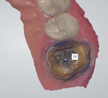 Fig. 11.3Magnified view of preparation
 -
Occlusal clearance indicated with color-coded ranges so that the dentist can evaluate the need for additional reduction (Fig. 11.4).
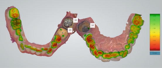 Fig. 11.4Occlusal clearance on prepared teeth
 -
Ability to verify virtual occlusion is identical to intraoral occlusion.
-
Preparation undercut detection (Fig. 11.5).
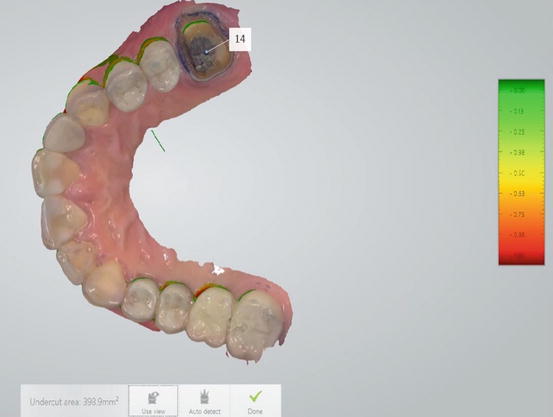 Fig. 11.5Undercut detection shown in green
 -
Ability to edit and modify impressions.
-
See chapter on Indirect Restorations with CAD/CAM Technology.
11.1.1.1 Advantages
-
Enhanced patient comfort
-
Efficient submission to lab virtually
-
Improved lab communication – ability to include pictures, mark margins, and annotate on digital impression
-
Retake impressions at no additional cost
-
Reduction of necessary components for implant impressions
11.1.1.2 Disadvantages
-
Learning curve associated with initial use
-
High initial investment and/or recurring additional fees (i.e., licensing fees/click fees)
11.2 Proper Technique for Imaging
- (a)
Avoid redundancy during scanning – increases accuracy and decreases file size (i.e., begin scanning occlusal surfaces on the most posterior tooth, moving to the anterior; rotate to capture the lingual surfaces and move from anterior to posterior; rotate over tooth to capture buccal surfaces and move anteriorly; end on an occlusal surface) (Fig. 11.6).


