(1)
Department of Pathology, Radboud University Nijmegen Medical Center, Nijmegen, The Netherlands
Abstract
As enamel consists for almost 100 % of minerals, it cannot withstand the decalcification needed to allow paraffin sectioning. Therefore, in general, it is not present anymore in the histologic sections, and so, the histopathologist usually will not be faced with problems on how to interpret changes in this tissue type. However, as gross inspection of submitted teeth before they are processed for histology will already yield useful data, alterations in this dental hard tissue will nevertheless be mentioned briefly from a practical point of view while acknowledging that the topic is much more complicated than depicted below.
Enamel
As enamel consists for almost 100 % of minerals, it cannot withstand the decalcification needed to allow paraffin sectioning. Therefore, in general, it is not present anymore in the histologic sections, and so, the histopathologist usually will not be faced with problems on how to interpret changes in this tissue type. However, as gross inspection of submitted teeth before they are processed for histology will already yield useful data, alterations in this dental hard tissue will nevertheless be mentioned briefly from a practical point of view while acknowledging that the topic is much more complicated than depicted below.
Amelogenesis Imperfecta
Amelogenesis imperfecta is a hereditary condition afflicting the tooth enamel. It is subdivided in a considerable number of conditions depending on the clinical appearance, the kind of disturbance and the pattern of inheritance that may be autosomal recessive, autosomal dominant or X linked. For practical purposes, amelogenesis imperfecta will be discussed to allow recognition of the various subtypes as defined by their gross appearance, teeth showing either abnormal enamel caps of normal hardness or enamel caps consisting of enamel that is too soft and discoloured. The first is called hypoplastic and the latter hypomineralised or hypomatured. The ambiguities arising from this phenotypic classification are demonstrated by the combined occurrence of both hypoplastic and hypomineralised amelogenesis imperfecta in a family also having taurodontism [1], and it has been proposed recently that a classification based on the mode of inheritance may be more appropriate [2–5]. The current description aims to provide a practically applicable guide.
As amelogenesis is a hereditary condition, it afflicts in principle all teeth or nearly all teeth although there may be variation in expression. Moreover, the defect may be associated with other dental anomalies such as disturbed form, calcification of the dental pulp and delayed eruption of the teeth [6]. Occasionally, the condition is associated with dental follicular hamartomas and gingival hyperplasia [7].
Amelogenesis Imperfecta, Hypoplastic
Amelogenesis imperfecta of the hypoplastic type is the result of a decreased amount of enamel matrix laid down during tooth formation. Therefore, the enamel cap does not acquire its normal thickness. As mineralisation is normal, the hardness of the remaining enamel layer may be normal. However, the reduced thickness of the enamel cap causes an abnormal crown form of the involved teeth. These externally visible abnormalities may vary from an almost absent enamel cap to only a few irregularities in an otherwise normally formed tooth. These irregularities have been classified as rough, pitted and grooved that may run vertically or horizontally (Fig. 5.1a, b).
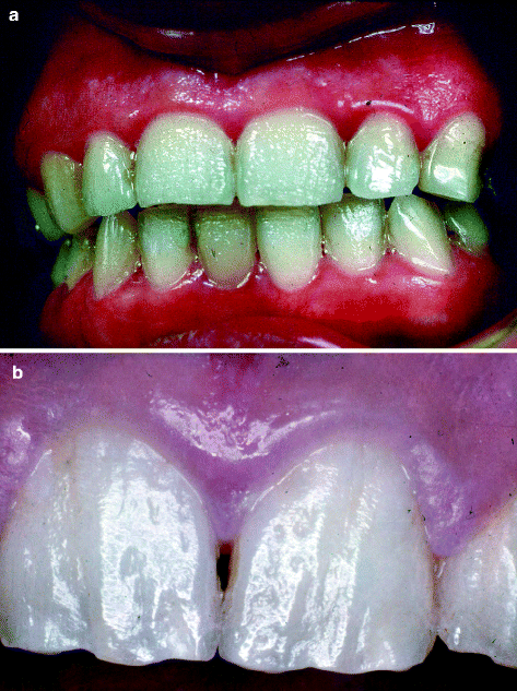
Fig. 5.1
Amelogenesis imperfecta of the hypoplastic type. Tooth surface shows pitting (a) or irregularities (b) indicating abnormal enamel formation. The hardness of the enamel is normal
Amelogenesis Imperfecta, Hypomineralised
In the hypomineralised type of amelogenesis imperfecta, the enamel initially develops a normal thickness, but the matrix is not mineralised in a normal way. Therefore, the teeth erupt with an initially normal appearance of their crowns, but the ill-mineralised enamel is soft and, therefore, is easily worn away in the mechanically demanding oral environment, thus exposing the underlying dentin. The enamel is light yellow-brown to orange and becomes brown to black after eruption because of stains from food or beverages. Characteristically, remnants of enamel remain present in mechanically privileged niches, for example, the cervical part of the tooth crown (Fig. 5.2a–d). Due to its low content of minerals, this defective enamel will not dissolve completely during decalcification, and therefore, it may be present in paraffin sections made from these teeth (Fig. 5.3a, b).
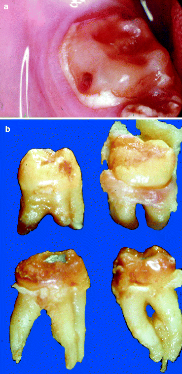
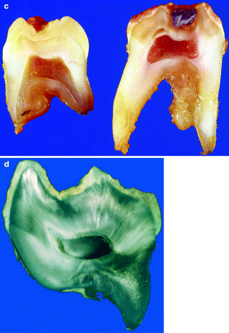
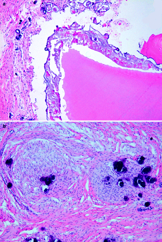
Fig. 5.2
Amelogenesis imperfecta of the hypomineralised type. The enamel cap is worn away due to masticatory forces leaving a bare dentin surface with enamel remnants present only at the cervical part of the crown (a). Gross appearance (b), cut surface (c) and ground section (d) showing thin and friable enamel cap
Fig. 5.3
Sometimes, teeth are covered by a defective enamel layer displaying both features of hypoplasia and hypomineralisation as shown in this photomicrograph. (a) The enamel layer shows variation in thickness and absence of regular lamellation (compare with Fig. 2.5). Underlying dentin does not show any abnormalities. (b) This case of amelogenesis imperfecta also showed enlarged dental follicles with areas of myxoid connective tissue in association with calcifications
Amelogenesis Imperfecta, Hypomatured
Amelogenesis of the hypomatured type is difficult to discern from the hypomineralised type as it also shows presence of enamel that is softer than it should be and that shows discoloration.
Enamel Hypoplasia
Enamel hypoplasia means any disturbance in tooth formation leading to macroscopically visible defects in the surface of the enamel. Although this definition also would include the hypoplastic form of amelogenesis imperfecta, the term enamel hypoplasia in practice is only applied to enamel lesions due to a systemic interference. This means that alterations may not be universal in that sense that they involve all teeth or the entire teeth crown as is the case in amelogenesis imperfecta. Only the enamel formed during the time span in which the systemic interfering factor was active will be abnormal. As the enamel is lost during decalcification, microscopy of this form of abnormal enamel requires ground sections which are not within the reach of most histopathology laboratories. However, macroscopical inspection of these teeth will already allow the distinction from amelogenesis imperfecta as in this latter disease; all teeth show abnormalities, whereas, in enamel hypoplasia, only some teeth may be abnormal. A common cause for enamel hypoplasia is an overdose of fluoride. This condition will be discussed more extensively below. Also tetracycline may give rise to enamel hypoplasia, but as this drug particularly causes tooth pigmentation, this topic will be discussed under that heading.
Enamel Opacities
Enamel opacities are white opaque spots in smooth-surfaced enamel. Probably they are due to transient hypomineralisation of the enamel matrix. Most often, they are seen at the incisor teeth (Fig. 5.4). Incidentally, they may also be brown and mottled. In that case, distinction from local enamel hypoplasia may become equivocal. The prevalence of this condition is rather high. Probably, enamel opacities are the result of short intervals of disturbed deposition of enamel matrix. Subsequent normal formation of enamel matrix will then bury the abnormal area.
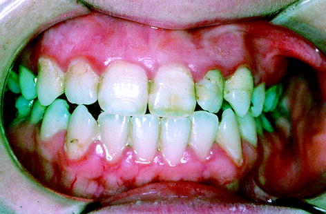
Fig. 5.4
White spot on labial surface of right first incisor tooth indicates transient period of hypocalcification
Dental Fluorosis
Fluorosis is a permanent hypomineralisation of enamel, in its mildest form characterised as small white spots. More severe forms range between white opaque areas to darkly stained and pitted enamel (Fig. 5.5a, b). Probably, an overdose of fluoride interferes both with function of ameloblasts and proper calcification of the enamel matrix [8, 9].
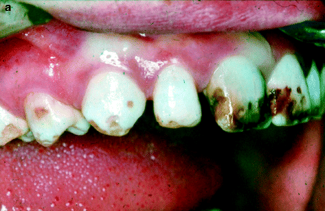
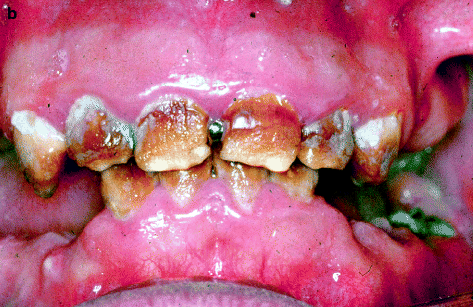
Fig. 5.5
Dental fluorosis. The abnormalities may vary from moderate (a) to severe (b). Enamel shows white spots, brown discolorations and surface irregularities indicating both enamel hypocalcification and hypomineralisation
Dentin
Structural abnormalities of dentin mostly have a hereditary background with an autosomal dominant way of transmission. In general, there is a failure of normal dentin formation after the initial deposition of a small amount of mantle dentin. As a result of this, involved teeth may show either abnormally short or entirely absent roots, whereas the pulpal chambers may obliterate partly or entirely due to formation of abnormal dentin masses. Two main groups are recognised: dentinogenesis imperfecta and dentinal dysplasia [1, 10–12].
Three different types of dentinogenesis imperfecta are recognised: type I that forms part of the spectrum of abnormalities that is shown by the generalised skeletal disorder osteogenesis imperfecta and types II and III that both occur isolated. Tooth crowns show an amber discoloration (Fig. 5.6) and have an ovoid shape, due to cervical constriction. Roots are short and thin. The pulp chambers are initially larger than normal but, with course of time, become almost obliterated by ongoing abnormal dentin formation. As the covering enamel tends to fracture from the underlying weakened dentin layer, the impression of coexisting amelogenesis imperfecta may arise [3, 4].
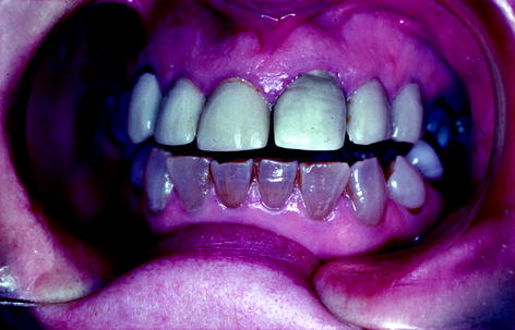
Fig. 5.6
Dentinogenesis imperfecta; teeth with amber discoloration (Picture kindly provided by Dr. I.G. Koutlas, Oral and Maxillofacial Pathology, School of Dentistry, University of Minnesota, Minneapolis, USA)
Dentinogenesis imperfecta type I and type II are clinically similar and only discernable by the already mentioned association of type I with osteogenesis imperfecta. Type III, which is limited to a specific racial subpopulation, differs from type I and II in not showing a time-dependent obliteration of the pulp chamber, teeth remaining extremely thin and therefore known as shell teeth (Figs. 5.7 and 5.8
Stay updated, free dental videos. Join our Telegram channel
VIDEdental - Online dental courses


