Crowns and fixed dentures
Orthodontic appliances
1a Crowns, 1b inlays/onlays, 1c bridges, 1d prostheses
2 Veneers
Removable dentures
Other custom–made products
3 Partial, 4 total
5 Splints, 6 epitheses etc.
15.7 Liability Risk from Dental Treatment
Treatment risks are very diverse and may lead to a liability of the practitioner (Gümpel 1994) (Table 15.1).
Table 15.1
Treatment risks (risk factors, patients at risk (Gümpel 1994))
|
Category 1
|
Category 2
|
Category 3
|
|---|---|---|
|
Somatization disorders
|
Dialysis patients
|
Hemophilia of various genesis
|
|
AIDS
|
Endocarditis hazard
|
Warfarin therapy
|
|
Blood disease
|
Endocrine disorders
|
|
|
Diabetes mellitus
|
Cardiovascular problems
|
|
|
Radiotherapy
|
Organ transplants
|
|
|
Cytostatic therapy
|
Pregnancy
|
15.7.1 Preliminary Comments
Anamnesis is of major importance before starting a treatment. The biggest mistake of the practitioner is to neglect the anamnesis, which requires not only general basic knowledge, but also some special expertise. This can lead to serious malpractice, as often shown in psychosomatic consultation, especially with patients with orofacial somatization disorders.
Marxkors (1999) describes a treatment that ended as a total failure. An otorhinolaryngologist referred (via telephone) a patient with a trigeminal neuralgia to his dental colleague. After hearing about the patient’s discomfort, the dentist immediately started an occlusal treatment in the right upper jaw. The treatment with four crowns in the mandible and a five-unit bridge in the maxilla was a failure. The bridge in the left upper jaw was renewed and the second copy, together with three crowns on the right, had to be removed and replaced by temporaries. During the treatment period, which extended over 9 months, the complaints were sometimes less, sometimes even unbearable.
If the dentist had read the medical history more carefully, he would have seen that the patient had more than 70 appointments during the last 2 years and a 10-year odyssey from specialists of internal medicine to holistic medicine. The patient himself described his discomfort in a previous medical history as “pain in various areas, depression and anxiety…” Gathering a complaint history is the first step and essential for all treatment sequences.
In view of the previous medical history, there was no chance to achieve success solely by dental treatment (Marxkors 1995; Marxkors and Wolowski 1999). Because no medical history was collected, there were no therapeutic–diagnostic investigations, no investigation of the quality of pain, no anesthesia test, no provocation test, no resilience test of the TMJ, and no instrumental functional analysis. This is forensically classified as culpable negligence, because basic knowledge of psychosomatic medicine is required. The statement that the complaints could not be reconciled with the findings raised in line would have led to the suspicion of an orofacial somatization disorder (Bräutigam et al. 1992).
15.8 Dental Procedures and Endocarditis—Prophylaxis
Patients with congenital heart defects are the main risk group for infectious endocarditis (Chart 15.2). Endocarditis prophylaxis is still insufficiently implemented in dental practice (Knirsch et al. 1999). Because of the pathogenesis of infective endocarditis (IE), the possibility of preventive measures in dental surgeries is given. To avoid any failure, dentists must be made aware of this while they are in training.
Chart 15.2
Graduation of the endocardium risk (Knirsch et al. 1999)
|
No increased risk:
|
|
Mitral valve prolapse without insufficiency noise
|
|
Condition after:
|
|
Coronary bypass operation
|
|
Pacemaker or defibrillator
|
|
Implantation of ventriculoperitoneal or ventriculoatrial shunts
|
|
Closure of ductus arteriosus (ductus Botalli)
|
|
Operated heart defects without residual findings (after the first year after surgery)
|
|
Isolated aortic isthmus stenosis
|
|
Atrial septal defect of secundum type (ASD II)
|
|
Increased risk:
|
|
Congenital heart defects (except atrial septal defect of secundum type (ASD II))
|
|
Acquired valvular heart disease
|
|
Postreeration heart defects with residual findings (without residual findings: only for 1 year)
|
|
Mitral valve prolapse with insufficiency noise without any distinctive myxomatous degeneration
|
|
High risk:
|
|
Heart valve replacement using mechanical or biological prostheses
|
|
Condition caused by microbial endocarditis
|
|
Congenital (complex) heart defects with cyanosis
|
Bacteremia caused by dental procedures can lead to microbial colonization (given predisposing endocarditis damage) (Horstkotte et al. 1997). After dental–surgical procedures with risk of bleeding, severe bacteremia often occurs, especially with dental extractions, intraligamentous anesthesia, dental cleanings, and in periodontal surgery, root canal treatment, and other dental surgical interventions. Because the oral cavity, under physiological conditions, is populated with more than 200 different species of bacteria and, in particular, the dental sulcus has a particularly high density of bacteria, a high quantity of bacteria reaches the predilection site, compared with other medical procedures (Knirsch et al. 1999).
Bacteremia after dental procedures usually does not last longer than 15 min after the causal surgery. Therefore, a single dose of an antibiotic is prophylactically sufficient. The proposed prophylaxis regimes are adequately tested in animal studies (Glauser and Bernhard 1983; Horstkotte 1985; 1995; Malinverni et al. 1988; Pippert et al. 1991). In the presence of a penicillin allergy, clindamycin provides an equivalent alternative (Table 15.2) in dental practice. This is in line with the recommendation of the American Heart Association (AHA) for high-risk patients. Amoxicillin per os (60 min before the planned operation) ensures a sufficiently high serum level to prevent the attachment of bacteria to the vegetation (Dajani et al. 1994).
Table 15.2
Orally applicable prophylaxis at dental surgeries (Horstkotte et al. 1997)
|
Child
|
Adult
|
|
|---|---|---|
|
Without penicillin allergy
|
50 mg/kg amoxicillin p.o. 60 min before surgery
|
2 g (<70 kg) to 3 g (=70 kg) amoxicillin p.o. 60 min before surgery
|
|
With penicillin allergy
|
15 mg/kg clindamycin p.o. 60 min before surgery
|
600 mg clindamycin p.o. 60 min before surgery
|
Martin et al. (1997) evaluated 53 cases of endocarditis that ended in court proceedings. In 48 of the 53 cases, no antibiotic was administered, in 2 cases it was either ineffective or administered at the wrong time. Only in one case was the antibiotic correctly administered. In a German Heart Centre Berlin (DHZB) retrospective analysis over a period of 10 years, endocarditis occurred with dental surgery as the supposed trigger in 4 of 22 patients with congenital heart disease (Knirsch et al. 1999).
15.9 Tooth Restoration Before and After Organ Transplantation
Dental, oral, and maxillofacial surgery is included into the preoperative and postoperative requirements, which ensure a lifelong immunosuppression to maintain the function of the transplant (Otten 1999). Patients with organ transplants have a higher risk for local or hematogenous and transmitted bacterial infection in the oral cavity because of the vital and lasting immunosuppressive medication.
The oral cavity has diverse bacterial colonizations under the predominance of a protective physiological streptococcal flora. In the majority of adult patients, pathogenic bacteria are also potentially present; they have their main location in the periodontium of the teeth. In addition to specific streptococcal species, there are mainly gram-negative anaerobic bacteria. Quality and quantity of the resident flora are not individually assessable, nor is the concrete pathogenic potency. Because of the resident flora of the mouth, the tooth can be an entry point of bacterial infections, manifested as an abscess or osteomyelitis.
The hematogenous spread of the potentially pathogenic agent mixture (known in endocarditis as oral streptococci) may lead to a focal dental infection (secondary bacterial infection). In immunosuppressed organ recipients, anaerobic pathogens (detectable in blood) also have a pathogenic significance.
Before transplantation, this difficulty requires:
1.
Clinical and radiographic (panoramic radiograph) investigation and documentation
2.
Hygiene instruction and professional tooth cleaning
3.
Conservative restoration (endodontics)
4.
Surgical rehabilitation
5.
Immediate prosthetic supply
6.
Reporting of completed dental restoration
Radical restoration treatments are not justified according to current knowledge. Every procedure must take into account the underlying disease and must be performed in consultation with the treating physician (in case of kidney dialysis before the transplantation, heart insufficiency before the heart transplantation, coagulation disorder before liver transplantation, diabetes before the pancreas transplantation, etc.). In individual cases, because of the risk of general complications, the limit of the outpatient treatment options can be exceeded. The fully completed dental restoration (including controlled wound healing) must be documented and reported. This communication is a prerequisite for the approval for the planned on-demand organ transplants.
Analogous to the current recommendations of the American Heart Association (Dajani et al. 1994), the following treatments have an increased risk for endocarditis:
1.
Tooth extraction
2.
Surgical removal of teeth
3.
Apicoectomy
4.
Periodontal treatment
5.
Periodontology (PAR) examination with probing or PAR surgery
6.
Tooth or implant cleaning with local bleeding
7.
Dental implant placement and reimplantation
8.
Endodontics with preparation
9.
Intraligamentous injection (local anesthesia)
15.10 Dental Treatment of Patients with Pacemakers
Patient must be asked whether they have a pacemaker. Patients with pacemakers are in possession of a pass that contains all relevant data about the pacemaker type and function, the pacing rate, the electrode type, etc. The pacemaker patient must not be seen as a “problem patient.” Routine questioning for medical history usually includes the reference of a pacemaker or implanted defibrillator (ICD).
15.11 Dental Surgery and Diabetes
In the Federal Republic of Germany, more than eight million people suffer from diabetes mellitus. Long-existing and inadequately controlled diabetes mellitus leads to microangiopathy and macroangiopathy, manifested as retinopathy, glomerular sclerosis, neuropathy, and premature arteriosclerosis. This results in an increased incidence of myocardial infarctions, strokes, kidney failure, etc.
In addition to the medical consequences of dental surgery, the general susceptibility to infections and for the oral area in particular are:
-
Accumulated periodontal abscesses
-
Defective wound healing after extractions
-
Persistent ulcerations
-
Gingival hyperplasia
If metabolic control is unclear, if the insulin dosage and administration are ambiguous, and before long-term operations, communication with the treating physician is necessary. It should be noted that stress (even the stress of a surgery) can affect the stability of the diabetic state, as well as local infections, trismus, and any fasting. Therefore, a quick change in the insulin dosage and continuous glucose monitoring by both doctor and patient may be necessary. (Wahl 1996). Caution: Children and young people are affected by type II diabetes (formerly named adult-onset diabetes) with increasing frequency. Blood sugar problems are possible, especially in overweight children, and if diabetes runs in the family (Vetter 1999).
15.12 Dental – Surgical Treatment of Patients Taking Warfarin
In the Federal Republic of Germany, approximately 150,000 patients use anticoagulants. Therefore, the treatment guidelines for patients who are taking warfarin should be known to each practitioner (Lechler and Pape 1996). For the prevention of thrombosis and embolism, patients at risk are treated with long-term anticoagulants. The indirect-acting anticoagulants are coumarin derivatives. They alter vitamin K metabolism. By reducing the active form of vitamin K, the synthesis of the coagulation factors II, VII, IX, and X as well as the inhibitors of protein C and protein S are reduced in the liver. Common compounds are warfarin, Falithrom (warfarin), and Coumadin (warfarin). In Germany, phenprocoumon is mainly used. Indications for anticoagulant therapy include vein thrombosis, pulmonary embolism, heart attack, atrial fibrillation, heart valve replacement, and certain heart valve defects.
Before dental–surgical procedures of patients who are taking warfarin, the dentist must be aware of the increased risk of bleeding (Fig. 15.1). The dimension of anticoagulation and thus the risk of bleeding is indicated by the prothrombin time (Quick) or variations such as the thrombotic test. The regular value for both tests is 100 %, with a normal range of about ±25 %. The therapeutic range, i.e., the range of the desired anticoagulation for the Quick test is between 15 and 25 %.
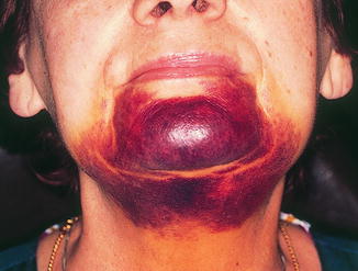

Fig. 15.1
Warfarin-treated patient. The interforaminal implantation was performed without taking into account the increased bleeding tendency
At extensive dental operations or dental restorations with insufficient possibility to stop local bleeding, a temporary increase of the Quick value to 30–40 % is suggested; from 1.6 to 1.9 INR (International Normalized Ratio). In cases of doubt, patients should be stationarily treated to reduce the risks of anticoagulants. In these cases, a syringe pump-controlled dose of heparin can eliminate the risk of thrombosis as well as short-term barriers to control the supply of heparin, intraoperative bleeding, or acute postoperative bleeding.
15.13 Treatment During Pregnancy
Pregnancy requires consideration of specific medical and legal concerns to avoid danger or damage to the unborn child. In addition, the physical and psychological characteristics of pregnant women should be kept in mind (Maeglin 1981).
15.14 Protection Against Radiation
The regulation of protection against damage caused by X-rays (§§ 22, 25, and 28) contains the specifics of ionizing radiation in women of childbearing age and during pregnancy. Although it is proven that prenatal exposure, depending on the dose and the gestational age, can result in death of the fetus, malformations, growth failure, malignant diseases, and genetic mutations, the radiation risk of dental X-ray images, in consideration of the optimal radiation protection, is extremely low. For shots of the mouth and jaw area, the radiation exposure for the uterus is estimated to be 0.1–1 GY.
However, X-ray examinations during pregnancy should only be performed if strictly indicated, in particular during the first period of gestation. To keep the radiation exposure as low as possible, highly sensitive films, rectangular tubes, and multiple X-ray protections are suggested. The number of shots must be kept to a minimum.
15.15 Toxicity of Amalgam Fillings
The Federal Institute for Drugs and Medical Devices currently recognizes no reasonable suspicion for embryotoxic or fetotoxic mercury risks from amalgam fillings during pregnancy, but recommends not to conduct further amalgam therapy during pregnancy and in women of childbearing age, because experimental, clinical, and epidemiological studies have shown that mercury is transplacentally transferred to the fetus.
The transfer of mercury from amalgam fillings by breast milk to infants has been demonstrated. A Swedish study of 30 mothers 6 weeks after delivery showed that there is a transfer of inorganic mercury from the blood into the milk, the mercury level in the milk was about half of that set by the WHO as the acceptable daily intake for adults (Oskarsson et al. 1996).
15.16 Prescription of Drugs
Even if prescription drug effects on the mother are known, the risk assessment of drug effects to the fetus is difficult. The nature of the fetal damage depends on the intrauterine development. The treating dentist is responsible for any drug therapy in pregnancy.
15.16.1 Antibiotics
Penicillin, cephalosporin, and macrolide antibiotics have no embryotoxic effects.
15.16.2 Analgesics
Taking into account some special features, the aniline derivative of paracetamol and antipyrine or propyphenazone is suitable for the use in pregnancy. Evidence of teratogenic effects are not available. Paracetamol crosses the placenta, therefore, high doses (for a longer time) should be avoided to prevent liver damage.
Aspirin (acetylsalicylic acid) inhibits prostaglandin synthesis and should therefore not be prescribed in the last month of pregnancy in order to prevent birth delay. Aspirin should not be given during pregnancy, because bleeding in pregnant women and fetuses were observed (aspirin may also lead to premature closure of the ductus arteriosus Botalli).
In special situations of pain with severe swelling, the use of derivatives of weak acids (nonsteroidal anti-inflammatory drugs) may be considered, such as arylacetic acids and arylpropionic acids; the best known are diclofenac and ibuprofen.
In special cases, the use of opiates and opioid analgesics, which are subject to narcotics regulation, is necessary. Evidence of a malformation potential is not found. In application shortly before birth, a respiratory depression of the newborn may occur. The long-term use of opiates can cause neonatal withdrawal symptoms and impairment of responsiveness.
15.16.3 Sedatives and Hypnotics
Anxiolytics may be prescribed for a short duration. The group of benzodiazepines is recommended because of the low rate of side effects. Diazepam is the most studied compound out of this group.
15.16.4 Local Anesthetics
Local anesthetics have a high lipid solubility and can therefore easily pass the placenta. The sooner the transition from maternal to fetal blood, the less the local anesthetic is bound to plasma proteins. Therefore, the local anesthetics with the highest protein binding rates are preferred. There are no reports of harmful effects to the fetus by local anesthetics during dental treatment.
Adrenaline as a vasoconstrictor additive is dosed as low as possible (1:200,000). On behalf of gynecologists, there are no objections to adrenaline derivatives, those substances are used as tocolytics (ache-inhibiting drugs). Intravascular administration must be avoided, because systemically resorbed adrenaline may lead to a constriction of the uterine vessels.
Norepinephrine and felypressin are contraindicated. Articaine, bupivacaine, and etidocaine may be used during pregnancy. The amides prilocaine and mepivacaine are to be considered more critically.
15.17 Damages Caused by Incorrect Treatment from the Perspective of Malpractice Assessments
Vogel presented (1982) a review of 584 claims, whereupon 42 % of dental damages were caused by surgical treatment (Tables 15.3 and 15.4), 32.4 % were caused by prosthetic measures (Table 15.5), and 16.6 % were caused by conservative treatments. Similar cases often resulted in widely divergent court decisions (Vogel 1982). Of medical liability lawsuits, 4 % are against dentists (Dajani et al. 1994). There is a comprehensive collection of judgments by Oehler (1999) concerning dental treatments and a list of information about various court judgments (BGH – Supreme Court, OLG – Court of Appeals, LG). Note also that the behavior after the occurrence of the damage will be assessed. A proper medical history, diagnosis, and treatment or referral to the appropriate further medical treatment is not only a medical duty but also a duty according to the insurance contract.
Table 15.3
Main complaints of 584 patients after dental treatment (Vogel 1982)
|
Damage caused by
|
n
|
Percent
|
From dental treatment
|
Percent
|
|---|---|---|---|---|
|
Surgery
|
246
|
42.1
|
10
|
4.0
|
|
Prosthetics
|
189
|
32.4
|
13
|
6.9
|
|
Conservative treatment
|
97
|
16.6
|
3
|
3.1
|
|
Local anesthesia
|
33
|
5.6
|
2
|
6.1
|
|
Orthodontics
|
15
|
2.6
|
1
|
6.7
|
|
General disease
|
3
|
0.5
|
1
|
33.3
|
|
General anesthesia
|
1
|
0.2
|
–
|
–
|
|
Total
|
584
|
100
|
30
|
5.1
|
Table 15.4
Liability claims after dental surgery (Vogel 1980a)
|
Caused by
|
Main accusation
|
Additional accusation
|
Justified
|
Compensation rejected
|
Unknown
|
|---|---|---|---|---|---|
|
Sensory disturbance
|
46
|
–
|
22
|
3
|
21
|
|
Untreated root residues
|
43
|
7
|
15
|
1
|
27
|
|
Broken jaw
|
33
|
–
|
12
|
4
|
17
|
|
Mouth–antrum contacts
|
24
|
–
|
6
|
7
|
11
|
|
Tooth damage
|
20
|
2
|
9
|
2
|
9
|
|
Confusion in extraction
|
19
|
–
|
11
|
1
|
7
|
|
Root fragments in the maxillary sinus
|
12
|
1
|
5
|
1
|
11
|
|
Untreated debris
|
12
|
1
|
5
|
1
|
6
|
|
Temporomandibular joint (TMJ) damage
|
7
|
–
|
0
|
1
|
6
|
|
Osteomyelitis, caused or incorrect treatment
|
6
|
3
|
2
|
2
|
2
|
|
Soft tissue injury
|
5
|
–
|
3
|
0
|
2
|
|
Incorrect diagnosis
|
4
|
–
|
1
|
2
|
1
|
|
Soft tissue infection, caused or incorrect treatment
|
3
|
–
|
0
|
3
|
0
|
|
Side confusion in operation
|
3
|
–
|
2
|
0
|
0
|
|
Lack of consent
|
3
|
–
|
2
|
0
|
1
|
|
Non-indicated extraction
|
3
|
–
|
0
|
2
|
1
|
|
Error in root tip resection
|
2
|
3
|
0
|
1
|
1
|
|
Preprosthetic surgery
|
1
|
–
|
0
|
1
|
0
|
|
Total
|
246
|
17
|
92
|
31
|
123
|
Table 15.5
Liability claims after conservative treatment (Vogel 1980b)
|
Caused by
|
Main accusation
|
Additional accusation
|
Justified
|
Compensation rejected
|
Unknown
|
|---|---|---|---|---|---|
|
Swallowing of root canal instruments
|
27
|
–
|
16
|
–
|
11
|
|
Soft tissue injuries
|
23
|
–
|
10
|
–
|
–
|
|
Broken root canal instruments
|
20
|
1
|
7
|
4
|
9
|
|
Via falsa
|
8
|
1
|
3
|
–
|
5
|
|
Damage after root canal treatment
|
6
|
–
|
1
|
2
|
1
|
|
Chemical burns caused by drugs
|
4
|
–
|
4
|
–
|
–
|
|
Incomplete root filling
|
3
|
–
|
–
|
2
|
1
|
|
Swallowing of filling parts
|
2
|
–
|
–
|
–
|
2
|
|
Inhaling of root canal instruments
|
2
|
–
|
2
|
–
|
–
|
|
TMJ dislocation
|
1
|
–
|
–
|
1
|
–
|
|
Secondary caries and pulp damage
|
1
|
3
|
–
|
–
|
1
|
|
Total
|
97
|
5
|
43
|
9
|
45
|
15.18 Dental Surgery
Klaus Rötzscher and4 and Rolf Singer5
(4)
German Academy of Forensic Odontostomatology (AKFOS), Wimphelingstraße 7, 67346 Speyer, Germany
(5)
Department of Oral and Craniomandibular Surgery, Clinicum Ludwigshafen/Rhein, Kalkofenweg 1, 67227 Frankenthal, Germany
Each oral surgical treatment bears complications that are either predictable or unpredictable (Egyedi 1998). Damages by surgical treatment measures are the most common complaints; nerve damage is the most common cause for liability claims (Vogel 1980a, 1982).
15.18.1 General and Local Anesthesia
If general anesthesia is performed by an anesthesiologist in dental practice (outpatient), the dentist is responsible for the indication of the anesthesia, but not for the implementation. The anesthesiologist is responsible for the method of anesthesia (Günther 1966).
Differentiated local anesthesia has been established as a fundamental form of analgesia in dental treatment of children as well as in adults. If, in certain situations, a treatment under local anesthesia is not considered possible by the dentist, or on the recommendation of the attending physician (GP/Internist), dental treatment may be carried out under general anesthesia. This may occur, for example, in acute diseases (e.g., inflammatory processes or trauma) and/or as a result of general medical risks or medical history (such as physical, mental, and psychological disabilities) or behavioral disorders. If the dentist comes to the conclusion that a further and adequate treatment under local anesthesia is not possible, e.g., unwilling children, this can also be seen as an indication for the implementation of a general anesthesia.
General anesthesia should only be performed by a doctor with the academic title “Doctor of Anesthesiology.” “The anesthesiologist is in charge for the anesthesia procedure and for monitoring of vital functions during and after surgery. This includes the management of complications and the incident involved therapy during and after anesthesia. After an outpatient’s anesthesia, monitoring to stabilize the vital signs is particularly important. The determination of timing and modalities of the home travel is one of the most responsible duties of care for anesthesia physicians” (Opinion of the German Association of Anaesthetists – In: Anesthesiology and Intensive Care Medicine 2/89).
In the decision between a nasal or oral intubation, the specific requirements of dental treatment are to be considered. With the invention of the intraligamental anesthesia procedure, the problem of reuse of opened cartridges arose, because this form of anesthesia often requires very little anesthetic and therefore (motivated by economy), one carpule is reused (with different needles) in other patients.
15.18.2 Risk of Infectiousness
Risk of even 1 % or less in view of the negligent transmission of infectious diseases is too high. The most important example is the risk of negligent transmission of hepatitis B (and HI) virus. Not only the injection needle but also the carpule must be replaced after each patient, even if anesthesia fluid is lost (Maeglin 1981). Disregarding this is a grossly negligent act, with all of the consequences of culpable behavior. “During injections of local anesthesia it may come to cardiac, central nervous and general reactions and side effects of the vasoconstrictive additives (especially in overdose, inadvertent intravascular injection and non-regarding of predisposing diseases, but also during local anesthesia performed lege artis). This may become acutely life-threatening and requires instant countermeasures” (Frenkel 1978).
Even a properly applied adjuvant (adrenaline) has a significant absorption rate in healthy patients (Lipp et al. 1988). Therefore, it is obvious that general intoxication can occur in correctly administered, and even more dramatically in accidentally administered, intravascular administration of catecholamines. Local anesthetics, accidentally injected intravenously with addition of a vasoconstrictor, have a significantly higher toxicity (Niesel and Kaiser 1991). Failure of aspiration followed by intravascular injection may lead to the accusation of breach of duty, similar to the accusation of disregarding previous illness or exceeding the maximum amount of supplementary injection.
It is a culpable act to leave (after local anesthetic) a pale and dizzy patient unattended. Less known are symptomatic psychoses in the sense of ordered semi-consciousness, presumably often misinterpreted because of their volatility, so the patient is not able to drive a motor vehicle for the next 24 h.
15.18.3 Infiltration Anesthesia and Fitness to Drive
A causal connection between infiltration anesthesia and a disturbance of consciousness (causing an accident) is often denied because of the low concentration of active ingredients and temporal relation. Both, however, can be explained by pharmacokinetics of the combination of local anesthetic and vasoconstrictive substance. The vasoconstrictive addition is intended to prevent a rapid absorption of local anesthetic at the target site and to prolong and deepen the effect. If the vasoconstrictive effect fades after 1–3 h, the vasodilatory effect of the local anesthetic in the well-vascularized mucosa starts. This can lead to increased washout and a short-term influx of the drug into the bloodstream. Local anesthetics are not metabolized at the site of action, but in the liver, some already partially in the blood. An effect similar to that of a relative overdose would be possible.
The absolute level of an active ingredient is less significant than the increase of concentration. In this context, it should be noted that clouding of consciousness, according to the severity classifications, can already be observed after a mild intoxication (Hempel et al. 1982).
Vasoconstrictors are added to local anesthetics to improve the local effect and to lower the blood levels, which are reduced after the absorption of local anesthetic as an expression of distribution of the substance in the body. The correctly administered local anesthetic in addition to a vasoconstrictor seems more compatible as a possibly vasoconstrictor-free substance. If the vasoconstrictor is injected intravenously by mistake, this increases not only the local anesthetic effect, but also the vasoconstriction (Niesel and Kaiser 1991).
There is a considerable link between oral infiltration anesthesia and the ability to drive. Conductive anesthesia must not be given if it is known that the patient will drive a motor vehicle after treatment. A limitation of driving ability may already arise from stress caused by dental surgery and must be part of the education. In addition, a duty of education also arises from relevant warnings by manufacturers of the anesthetics. According to current Supreme Court jurisprudence (BGH), the physician must inform the patient about all possible risks that cannot be excluded with certainty (Haffner and Graw 1995).
15.18.4 Postoperative Sensory Disturbances
Irreversible sensory loss after dental treatment (mainly after tooth extractions and surgical procedures, less after anesthesia) has significantly increased in recent years (Vogel 1980a, b). Contact of the root tip to the mandibular canal or a direct trauma by instruments or dislocated roots can result in irreversible damage to the N. alveolaris inferior.
The N. mandibularis (Figs. 15.2 and 15.3) is a mixed but predominantly sensory nerve. It reaches the infratemporal spatium through the foramen ovale. The locomotor units of the chewing muscles leave the nerve trunk in the infratemporal fossa. Afterwards, the bifurcation distributes in sensory branches (Evers 1983). Prior to each extraction, an X-ray image should be taken. In order to detect rare anatomical variations, the dentist must be oriented to the anatomic conditions in the root zone in order to identify the nerve course (Figs. 15.4, 15.5 and 15.6). The N. lingualis is usually injured by slipping instruments in the floor of the mouth or by conductive anesthesia at the wrong place. Scoring the periosteum with the needle tip during injection must be strictly avoided (Fig. 15.7).
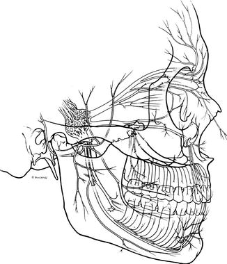
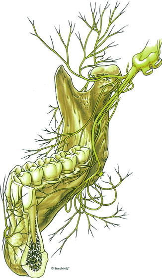
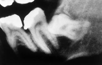
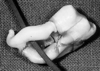
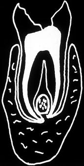
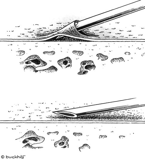

Fig. 15.2
N. mandibularis, side view (Evers 1983)

Fig. 15.3
N. mandibularis, frontal view (Evers 1983)

Fig. 15.4
N. mandibularis, X-ray before extraction (operation)

Fig. 15.5
N. mandibularis, presentation of the nerve course

Fig. 15.6
N. mandibularis, presentation of the course of the nerve (drawing)

Fig. 15.7
The needle tip should never be introduced into the foramen, because the risk of vascular or nerve injury is high and complications such as hematoma and paresthesias (longer lasting) can occur (Evers 1983)
15.18.4.1 Types of Nerve Lesions/Complications
Injury to nerves, arteries, or veins during the local/conductive anesthesia may be caused by:
-
The surgical procedure itself
-
Contaminated needles with subsequent infection
-
Quick injection with subsequent postoperative pain by ripping the tissue or necrosis
-
Large injection volume (do not use more local anesthetic than necessary) or injection into infected area
-
Trismus (usually caused by injection into the M. pterygoid internus)
-
Bleeding at the site of injection (using solutions with low-dosed vasoconstrictor additives)
-
Facial paralysis (injection at the posterior border of the ascending ramus) with atony of the lower lip (hanging mouth) (Evers 1983)
15.18.4.2 Consequences of Nerve Lesions
Typical clinical symptoms of nerve injury are:
-
Hyperesthesia
-
Hypoesthesia
-
Paresthesia
-
Anesthesia and anesthesia dolorosa
15.18.4.3 Duration of the Nerve Injury
Lesions may be temporary or permanent (>2–3 years after surgery). Injuries of the sensory branches of the N. trigeminus are a very painful complication. Most common are impairments of N. alveolaris inferior, the N. mentalis, and the N. lingual (Opinion DGZMK 6/ 89). With the increasing awareness of modern microsurgical nerve–surgical options to restore function after nerve lesions, considerable progress has been achieved. Damages to the nerves are no longer accepted as an irreversible fate, because patients are now referred to microsurgical therapy at an early stage.
In cases where there is a close anatomic relationship of N. lingualis to the mandible body and N. alveolaris inferior to the teeth, there is a risk of damaging those nerves during dental, surgical, and some conservation measures. The most common lesion sites of the N. lingual and alveolaris inferior are in the range of the mandibular angle, especially in the third molar region.
According to OLG Munich (Higher Regional Court), Judgement of 20.11.1996, Az. 4810 AHRS, “Prior to conductive anesthesia, an education about the rare and usually avoidable risk of damaging the N. Alveolaris is not required.” In contrast, the OLG (Higher Regional Court) of Munich ruled (Az. 26–O–285/78) with the tenor that it is a rare (0.1 %) but typical complication. The N. Mentalis is mostly injured outside the mental foramen (injury of branches of the trigeminal nerve II are very rare). Nerve injuries are usually not noticed during the operation. The often-described symptom of a dull pain occurs only rarely.
15.18.4.4 Symptoms of Nerve Injury During Injection – The Immediate Pain
If the trunk of the N. lingualis or N. mandibularis is hit during conductive anesthesia, this is accompanied by a flashing defensive movement of the patient or with a shooting pain in the lower lip or tongue. This phenomenon always should be documented in the patient file in order to point to a possibly sensory disturbance.
If, as a result of close anatomical relationship of the root tips to the mandibular canal, the risk was obvious, a claim for compensation may be entitled because of lack of education (lack of preoperative X-ray diagnosis or incomplete image of the root tips in case of damage). A survey (during a legal dispute) showed that a N. lingualis injury in a sole anesthetic block may occur only by the pressure rise caused be the anesthetic solution, or more likely caused by hemorrhage and clot formation and subsequent fibrosis in and around the nerve.
15.18.4.5 Injury of the N. Mandibularis During Surgery
Prior to the surgery, the dentist must inform the patient about the risk of irritation of the nerve. There is current consensus that, in a nonurgent medical intervention, the rare complication of nerve injury must be explained to the patient (OLG Köln, Judgement of 22.8.1998, Az. 5 U 232/96). LG Dortmund, Judgement of 27.09.1979, Az 2O29/79; OLG Hamm, Judgement of 12.02.1980, Az AHRS 4800/1; LG Mannheim, Judgement of 04/30/1986, Ref 9 – O-6/85; OLG Dusseldorf Higher Regional Court, Judgement of 25.07.1991, Az. AHRS 2694/12 states that “Prior to a non-critical extraction/surgical removal of a wisdom tooth, the patient must be educated about the risk of damage to the N. mandibularis and its consequences.”
Very often the clinician tries to shift the responsibility for an injury of N. mandibular to a postoperative tissue swelling or a postoperative bleeding. OLG Dusseldorf Higher Regional Court, Judgement of 25.07.1991, Az. AHRS 2694/12, and OLG Stuttgart Higher Regional Court, Judgement, Az. 12 U 25/95 found that “The postoperative tissue swelling is not such as the nerve (covered by a bony layer) gets damaged. It is also possible that the nerve injury is based on a post-operative bleeding, because bleeding caused by such compression, leads only to temporary neural deficits.” The dentist’s argument that a scarred structure around the nerves was the cause of the injury was dismissed by court because the sensitivity loss had occurred immediately after surgery but a scar formation takes days or weeks.
Damage to the N. mandibularis can also occur during an implantation in the mandible. For example, during the postoperative control after an implantation in the left and right lower jaw, the patient complained about numbness in the N. mandibularis. A CT scan was recommended (Figs. 15.8 and 15.9). The family dentist removed the two implants and replaced them by 2-mm-shorter implants. The paresthesia was completely healed within 3 months.
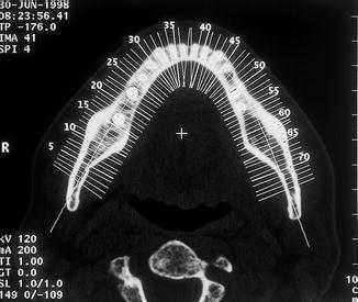
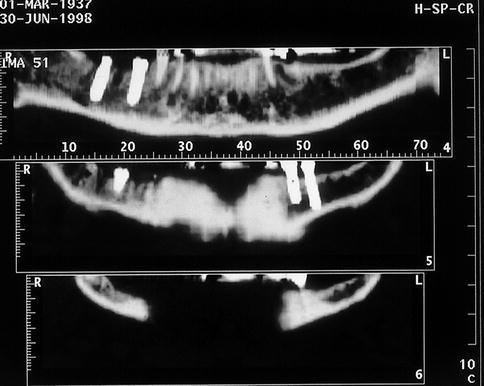

Fig. 15.8
CT scanning

Fig. 15.9
Both implants in the right and left mandible are displayed
In postoperative lesions/irritations of the N. mandibularis after implantation, a three-dimensional image should be taken to discuss the therapeutic alternatives with the patient. Both distal ends (47 and also 46) project themselves into the nerve canal (Fig. 15.10). The radiograph (see Fig. 15.9) (layer 54) shows that the drill hole passes lingually.
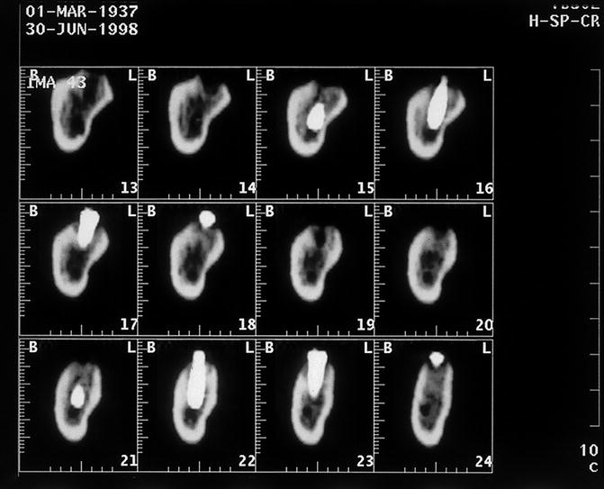

Fig. 15.10
Both distal ends (47 and also 46) project themselves into the nerve canal
A lesion of the N. mandibularis may be related to a root filling in the lower posterior teeth by the passage of the root canal in the mandibular canal. For example, the distal implant (coating level 54) is projected above the nerve canal (Fig. 15.11). The X-ray control must be performed immediately after root canal filling to detect the passage of the material into the nerve region in time (Fig. 15.12). Thus, the very different toxicity of the root canal fillings has a significant role. The panoramic display shows the expansion of the overstuffed root filling. Contrary to primary advice, an intraoral neurolysis was conducted to remove the root filling material. This resulted in a rupture of the nerve, which was followed by a nerve transplant and ended up in anesthesia dolorosa.
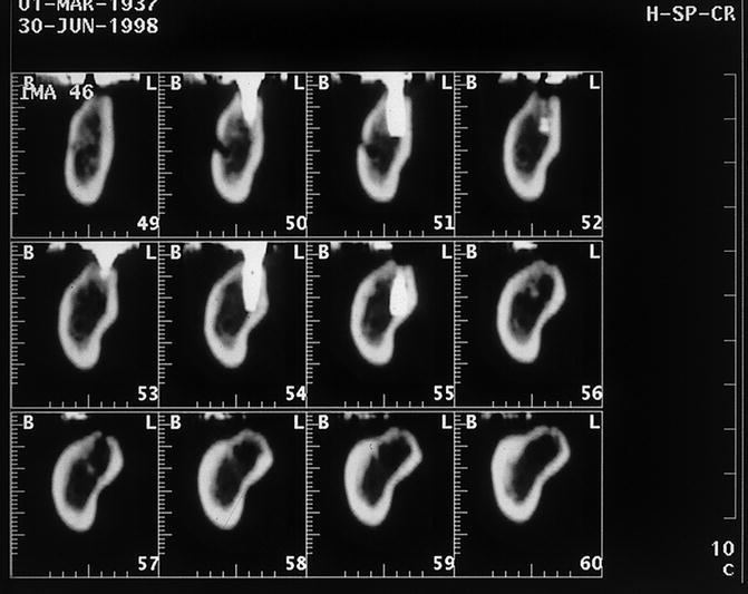
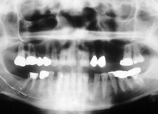

Fig. 15.11
The distal implant (coating level 54) is projected above the nerve canal

Fig. 15.12
Condition after root treatment of tooth 46 with overstuffed and spread filling material
Chloropercha, e.g., develops relatively low toxic stimuli compared with compounds that include plastic state unpolymerized constituents or iodoform or paraformaldehyde. Additionally, calcium hydroxide preparations are not harmless, because they are highly alkaline and etching. Toxic damage occurs immediately and the material must be removed quickly. Because root canal filling materials (once applied into the mandibular canal) must be removed by microsurgical interventions, it is wrong to extract the tooth (after overstuffing) with the intent that root filling material could be drained.
15.18.4.6 Injury of the N. lingualis During Surgery
Nerve injuries of N. lingualis can lead to numbness of the affected half of the tongue and/or to a unilateral failure of the taste perception. If only the sensitivity in the branching of N. Lingualis is disturbed, this indicates a partial lesion of N. Lingualis.
LG Frankfurt, Judgement of 02/03/1981, File number: 2/16 S 172/80 found that:
-
1. A nerve injury of the N. lingualis with permanent numbness of the left half of the tongue during the surgical removal of the impacted wisdom tooth 38 can not be causing for an infraction during the anesthesia or the surgery itself.
-
2. In approximately ten million cases of conductive anesthesia per year, only about ten lead to a long-term damage of the N. lingualis, so there is no duty to inform.
Stay updated, free dental videos. Join our Telegram channel

VIDEdental - Online dental courses


