Introduction
The aim of this study was to compare dental development in a group of children with mild-to-moderate hypodontia with a matched group.
Methods
A study group of 70 children (43 girls, 27 boys) with hypodontia, aged 5.3 to 12.5 years, was matched for race, age, and sex with 140 healthy, normal control subjects. The children’s dental ages were calculated by using a modified dental-age estimation method. Differences between dental and chronologic ages were analyzed by t tests, and the correlations between differences in dental and chronologic ages and the numbers of missing teeth were analyzed with the Spearman correlation test.
Results
Dental development in children with mild-to-moderate hypodontia was significantly delayed compared with the control group ( P <0.05); however, the mean difference did not exceed 0.3 years in either sex. No correlation was observed between the differences in dental and chronologic age and the severity of hypodontia.
Conclusions
Children with mild-to-moderate hypodontia had delayed dental development of a few months; this was statistically significant. Because of individual variations, each patient should be carefully examined.
One of the most common polymorphisms is hypodontia, the absence of at least 1 tooth, excluding the third molar. The prevalence of hypodontia in the permanent dentition has been reported to be 0.3% to 10.1%, depending on the population studied. Hypodontia is classified according to the severity of the condition, with “mild-to-moderate hypodontia” used to denote the absence of 2 to 5 teeth and “severe hypodontia” denoting the absence of 6 or more teeth.
Early diagnosis and effective clinical management of hypodontia are important because the condition can lead to esthetic, physiologic, and functional problems such as malocclusion, periodontal damage, and inhibited alveolar growth. The management of absent teeth generally involves a multidisciplinary approach. The number, size, and development of the remaining teeth are significant factors in long-term treatment planning.
Hypodontia has been reported to be associated with other dental abnormalities, including microdontia, transpositions, and taurodontism. Hypodontia can also be associated with delayed tooth formation ; however, there is no consensus in the literature concerning this latter phenomenon.
In this retrospective, cross-sectional study, we aimed to compare dental development in a group of children with mild-to-moderate hypodontia and a matched group and to determine whether the severity of the hypodontia has an effect on dental development.
Material and methods
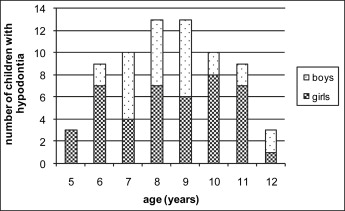
Hypodontia subjects were selected based on radiographs of patients who previously applied to the Department of Pediatric Dentistry at the Ondokuz Mayis University Faculty of Dentistry in Samsun, Turkey. In most cases, the patients were referred to the pediatric dental clinic because of caries-related pain or traumatic dental injuries. Patients with developmental anomalies such as ectodermal dysplasia, cleft lip or palate, and Down syndrome, as well as patients who had undergone previous orthodontic treatment, were excluded from the study. In previous studies, panoramic radiograph examinations were found to be a reliable method of diagnosing hypodontia. In this study, a tooth was diagnosed as congenitally missing when no mineralization of its crown could be identified on the panoramic radiographs, with no evidence of its having been extracted. Third molars were excluded. A total of 70 subjects (43 girls, 27 boys) were identified as having mild-to-moderate hypodontia ( Table I ). Table II shows the distribution of missing teeth according to age and sex. The mean chronologic age of the patients at the panoramic examinations was 8.9 years (range, 5.3-12.5 years) ( Fig 1 ).
| Missing teeth (n) | Girls (n) | Boys (n) | Total (n) |
|---|---|---|---|
| 1 | 18 | 11 | 29 |
| 2 | 16 | 10 | 26 |
| 3 | 3 | 2 | 5 |
| 4 | 5 | 3 | 8 |
| 5 | 1 | 1 | 2 |
| 43 | 27 | 70 |
| Age(y) | Sex | Missing teeth (n) | ||||
|---|---|---|---|---|---|---|
| Maxillary lateral incisor | Maxillary second premolar | Mandibular central incisor | Mandibular lateral incisor | Mandibular second premolar | ||
| 5.0-5.9 | Girls | 2 | 1 | 2 | ||
| Boys | ||||||
| 6.0-6.9 | Girls | 3 | 5 | 2 | 8 | |
| Boys | 1 | 2 | 1 | |||
| 7.0-7.9 | Girls | 2 | 2 | |||
| Boys | 6 | 6 | ||||
| 8.0-8.9 | Girls | 3 | 3 | 5 | ||
| Boys | 6 | 3 | 8 | |||
| 9.0-9.9 | Girls | 3 | 3 | 8 | ||
| Boys | 11 | |||||
| 10.0-10.9 | Girls | 5 | 4 | 6 | ||
| Boys | 2 | 1 | ||||
| 11.0-11.9 | Girls | 5 | 3 | 1 | 4 | |
| Boys | 2 | 3 | ||||
| 12.0-12.9 | Girls | 2 | 2 | |||
| Boys | 2 | |||||
| Totals | 42 | 25 | 2 | 5 | 64 | |
For every hypodontia subject (n = 70), 2 normal, healthy subjects matched for sex and age at the panoramic examination were selected at random from the Faculty of Dentistry records as control subjects (n = 140; 86 girls, 54 boys). The criteria for inclusion in the sample were the availability of a panoramic radiograph of adequate quality and no history of surgery or disease that could affect the development of the mandibular permanent teeth.
Chronologic age (CA) for each subject was calculated by subtracting the date of the panoramic radiograph from the date of birth after converting both to decimal ages.
The mandibular left canines, first premolars, and second molars in the children in both the hypodontia and control groups were scored according to the method of Demirjian et al, in which the formation of mandibular permanent tooth crowns and roots are divided into 8 stages, with detailed written criteria and supplementary illustrations provided for each stage. The scores were then correlated with median-age values identified in a previous study describing the chronology of mandibular permanent tooth mineralization in children in northern Turkey in line with the scoring system of Demirjian et al. In our study, dental age (DA) was calculated by adding the median-age values of the mandibular left canine, first premolar, and second molar and dividing by 3 (ie, the number of teeth scored).
The dental development of the study sample was assessed by a trained, blinded examiner (A.E.K.). The assessments were performed in a darkened room by using a radiographic illuminator to provide contrast enhancement of the dental images.
Statistical analysis
The Kolmogorov-Smirnov test was used to evaluate the normal distribution of variables. The t test was used to compare the mean differences in DA and CA (DA-CA) between the hypodontia and the control groups. The Spearman rank correlation analysis was performed to determinate the relationship between the severity of hypodontia (number of missing teeth) and the DA-CA values.
Intraobserver error was calculated by rescoring 20 randomly selected radiographs (60 teeth) 2 weeks after the initial assessments. Paired t tests were used to identify differences in DA-CA between the 2 readings. Agreement rates for the 2 readings were calculated, and bias was assessed by counting the numbers of over- and underassessments in DA between the 2 readings.
Stay updated, free dental videos. Join our Telegram channel
VIDEdental - Online dental courses


