Introduction
The objectives of this study were to clarify the characteristics of cranial-base morphology in adults with skeletal Class III malocclusion and investigate factors relating to the establishment of a skeletal Class III malocclusion.
Methods
Initial lateral cephalograms of women were examined. Subjects with an ANB angle of 0° to 4°, normal overjet and overbite, and a Class I molar relationship were classified as Class I (n = 86). Those with an ANB angle less than −1°, a Wits appraisal less than 2 mm, a negative overjet, and a Class III molar relationship were the Class III group (n = 86) in this study. Angular, linear, and coordinate measurements were made. Multivariate analysis of variance and the Student t test were used to analyze significant differences between the 2 groups. Discriminant analysis, logistic regression analysis, and decision analysis were used to identify which cranial-base and maxillomandibular variables influenced the establishment of a skeletal Class III malocclusion.
Results
The Class III group had smaller values for NSBa, SeSBa, FH-SSe, and FH-SBa. Sphenoidale and basion were more inferior and anterior than those of the Class I group. There was no difference in the anterior and posterior cranial-base lengths between the groups. Greater mandibular length was the first major characteristic in the Class III group, followed by smaller values for SeSBa and NSBa.
Conclusions
Cranial-base morphology in adults with a skeletal Class III malocclusion is different from that in a skeletal Class I malocclusion. Smaller cranial-base angles, steeper posterior cranial bases, more inferiorly positioned sphenoidale, and more anteriorly positioned basion are major characteristics of skeletal Class III malocclusions. These characteristics play important roles in the establishment of a skeletal Class III malocclusion.
A skeletal Class III malocclusion results from morphologic or positional disharmony between the maxilla and the mandible during the growth period. In 1916, Young reported that the cranial-base morphology might be related to jaw prognathism. Since then, several studies have investigated the relationship between the cranial-base and maxillomandibular components.
The cranial base includes both the anterior and posterior cranial bases. The anterior cranial base relates to the position of the maxilla, whereas the posterior cranial base relates to the positions of the glenoid fossa and the mandible. Recently, the correlations between the lateral basicranium and facial form, mandibular morphology and position, and malocclusion patterns have also been investigated. Bastir et al and Bastir and Rosas demonstrated that both the midline cranial base and the middle cranial fossa correlate with mandibular ramus morphology.
One of the most commonly used cranial-base characteristics is the cranial-base angle from the flexion of the anterior and posterior cranial bases at sella turcica in the midsagittal plane. It has been considered that the cranial-base angle has an influence on the anteroposterior intermaxillary relationship and the type of malocclusion. Furthermore, the dimensions of the cranial base play an important role in determining the sagittal jaw relationship.
A small cranial-base angle (NSBa) and a short cranial-base length (S-N) are major morphologic features of skeletal Class III patients. A deficiency of the cranial base that flattens during growth has been considered a factor associated with skeletal Class III malocclusion. However, recent studies have reported no correlation between the cranial-base angle and a skeletal Class III malocclusion.
Since the evidence is still inconclusive, whether the cranial-base morphology in a skeletal Class III malocclusion differs from that of a skeletal Class I malocclusion and has a relationship to the establishment of a skeletal Class III malocclusion remains controversial. Furthermore, most previous researchers selected their subjects irrespective of sex and age. There have been few cranial-base studies in adults with a skeletal Class III malocclusion.
The objectives of this study were to clarify the characteristics of cranial-base morphology in adults with a skeletal Class III malocclusion by comparing them with those with a skeletal Class I malocclusion, and to investigate factors related to the establishment of a Class III malocclusion.
Material and methods
Initial lateral cephalometric radiographs of 424 Japanese women, ages 16 to 35 years, from the Department of Orthodontics, Dental Hospital, of Tsurumi University in Yokohama, Japan, were examined in our study. The study protocol was approved by the institutional review board of the university. The image magnification of the cephalostat for this study was 10%, and all linear measurements were adjusted accordingly. All subjects were adults because all cephalograms showed stage 6 of cervical vertebral maturation, indicating that at least 2 years had passed after the pubertal growth peak. Subjects with an ANB angle from 0° to 4° with normal overjet and overbite, a Class I molar relationship, and a normal facial profile were classified as the Class I group, and those with an ANB angle less than −1°, a Wits appraisal less than −2 mm, a negative overjet, a Class III molar relationship, and a concave facial profile were the Class III group. The exclusion criteria were cleft lip and palate, craniofacial syndrome, mandibular deviation more than 5 mm, remaining deciduous or missing teeth (except third molars), a previous history of orthodontic treatment, and poor-quality cephalograms. Consequently, 86 skeletal Class I and 86 Class III subjects met the inclusion criteria. The skeletal Class I and Class III subjects’ ages ranged from 16.1 to 34.0 years (mean, 21.6 years; standard deviation [SD], 3.9) to 16.0 to 35.0 years (mean, 22.0 years; SD, 4.3), respectively.
Each lateral cephalogram was traced on 0.003-in frosted acetate by 1 investigator (S.S.) and checked for accuracy by another investigator (T.S.). Cephalometric landmarks were identified, digitized, and analyzed with CephaloMetrics AtoZ software (version 9.5J; Yasunaga Computer Systems, Fukui, Japan). Definitions for these landmarks are given in Figure 1 .
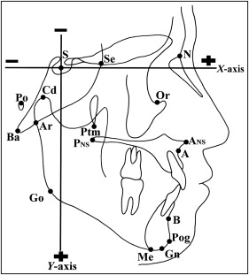
The positions of the landmarks were recorded in Cartesian coordinates (x- and y-axes) with sella as the origin of the axes (x, y = 0, 0). The horizontal reference plane (x-axis) was parallel to the Frankfort horizontal plane passing through sella. The vertical plane (y-axis) was set perpendicular to the horizontal plane. The anterior and inferior areas near sella were set to positive, whereas the posterior and superior areas near sella were set to negative.
To test the error of point identification, the lateral cephalograms of 25 subjects were randomly selected, retraced, and redigitized 2 months after the initial analysis by the same investigator (S.S.). Errors in identifying and locating cephalometric landmarks were assessed by Dahlberg’s formula (measurement error estimation), and the coefficient of reliability was calculated as follows.
where d is the mean difference between repeated measurements, and n is the number of measurements.
where S e 2 is the variance due to random error, and S t 2 is the total variance of the measurements.
Dahlberg’s errors were 0.17° to 0.50°, 0.14 to 0.39 mm, and 0.15 to 0.32 mm for angular, linear, and coordinated value measurements, respectively. The coefficient of reliability indicated that the observed values of the measured variables were between 0.925 and 0.999.
Statistical analysis
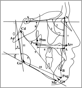
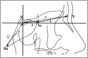
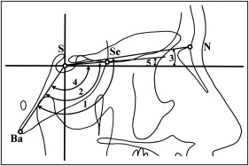
Angular, linear, and coordinate values measurements for groups of maxillomandibular variables and cranial-base variables were performed ( Figs 2-4 ). Significant differences between the Class I and Class III groups in maxillomandibular and cranial-base variables were tested with the Hotelling T 2 test (multivariate analysis of variance; MANOVA) as an initial exploratory test. When significance was found, the independent-sample Student t test was used to identify significant between-group differences for each cephalometric variable. Because of multiple testing, we adjusted the level of significance according to Holm’s sequentially rejective multiple test procedure.
Three statistical models—decision analysis by classification and regression trees, discriminant analysis by stepwise regression, and logistic regression analysis by forward selection—were performed to identify the variables in the cranial base, maxilla, and mandible that characterize skeletal Class I and Class III subjects. The independent variables were ramus inclination, mandibular plane angle, gonial angle, A′-Ptm′, A Mx H, P Mx H, Gn-Cd, Pog-Go, Cd-Go, NSBa, SeSBa, FH-SN, FH-SSe, FH-SBa, S-N, S-Se, N-Se, and S-Ba. To compare the accuracy of the 3 statistical models, the cutoff values were obtained from the receiver operating characteristic curves, and classification accuracy was called the crude hit rate. It was calculated as follows.
Crude hit rate = number of true positives + true negatives total number of subjects
Stay updated, free dental videos. Join our Telegram channel
VIDEdental - Online dental courses


