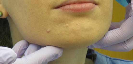
Fig. 1.1
Forehead and hairline (Photos courtesy of Anna Christine Napoli and Dr. Karin Schey)
-
Standing in front of the patient, visually inspect the face.
-
Look for symmetry; color, pigmentation, contour, consistency, and function.
Forehead and eyes
-
Palpate the forehead. Look for nodules, swellings, and masses (Fig.1.2).
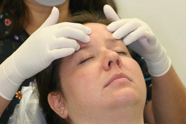 Fig. 1.2Palpation is a quick way to examine a patient for asymmetries
Cranial nerves and facial muscles
-
Have patient follow your fingers in an H pattern. Assess possible deficiency in facial nerve function (Fig.1.3).
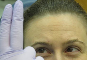 Fig. 1.3The patient should follow finger movements with the eyes, while not moving the head. Movement of the eye is controlled by the oculomotor, trochlear, and abducens nerves
 -
Have patient squint, look for symmetry. Asymmetry during squinting could indicate facial muscle deficiency (Fig.1.4).
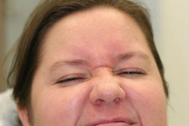 Fig. 1.4Squinting may detect facial nerve symmetry (normal finding) or asymmetry (abnormal finding)
Ears
-
Inspect and palpate all visible portions of the ear (Fig.1.5).
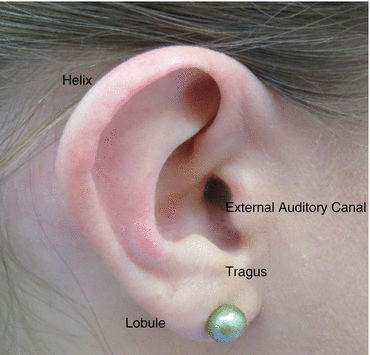 Fig. 1.5Outer ear
 -
Look for color, pigmentation, contour, consistency, and function.
Eyes
-
Inspect the eyes (Fig.1.6).
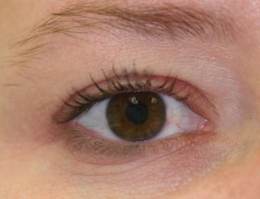 Fig. 1.6Check the eyes for sclera coloring, drainage, or swelling
 -
Look for white sclera, absence of swelling, or drainage.
-
Yellow sclera may suggest jaundice.
-
Hematoma could indicate a bleeding disorder or injury.
Nose
-
Look up the nares (nostrils) and palpate the nose (Fig.1.7).
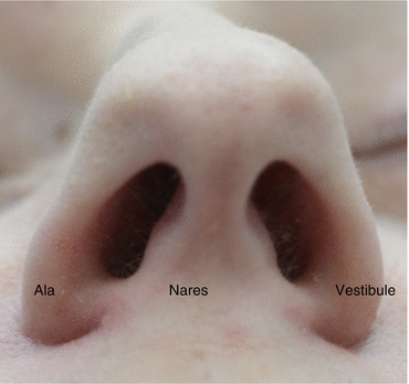 Fig. 1.7Lack of symmetry of the nose should be noted when performing esthetic dental procedures as it may be critical when determining facial midlines
 -
Look for nodules, swellings, and masses.
TMJ
-
Sitting or standing behind the patient, visually inspect the face. Look for symmetry in function. Note any abnormal deviation of the mandible.
-
Palpate the temporomandibular joint (TMJ) while having patient open and close with fingers placed over the condyles, bilaterally. Note clicking, popping, and discomfort/pain (Fig.1.8).
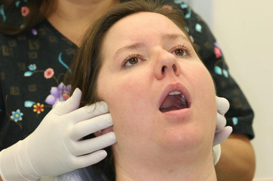 Fig. 1.8If TMJ symptoms are present, a more extensive evaluation will be necessary
 -
Look for nodules, swellings, and/or masses.
Parotid gland and preauricular nodes
-
Feel the parotid bilaterally and the preauricular nodes (Fig.1.9).
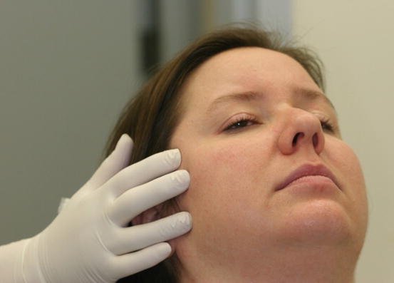 Fig. 1.9Bilateral palpation of the parotid gland and preauricular area
 -
Compare for symmetry, identify nodes by size and if they are hard or soft, painful or painless, and freely movable or fixed.
-
Possible findings: Lymph nodes may have nodules, swelling, and/or masses.
Posterior neck nodes
-
To palpate the posterior auricular and occipital nodes, drop head forward to enhance access to these areas.
-
Palpate over the trapezius muscle for the spinal accessory and posterior cervical nodes (Fig. 1.10a, b).
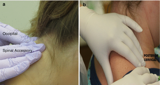 Fig. 1.10(a, b) Tenderness to palpation should also be noted
 -
Compare for symmetry and identify nodes for size, consistency (hard or soft), level of pain, or if freely movable or fixed.
Anterior neck nodes
-
Palpate the jugular chain, deep and superficial cervical nodes, by placing fingers firmly on both sides of the sternocleidomastoid muscle from its origin at the clavicle to its insertion at the mastoid process behind the ear (Fig. 1.11).
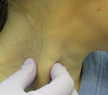 Fig. 1.11Jugular lymph node enlargement along the sternocleidomastoid is common in patients experiencing the common cold or with head and neck cancer (Photo Courtesy of Dr. Eugenia Monaghan)
 -
Palpate the supraclavicular, anterior scalene, and delphian nodes above the clavicles and near the inferior midline of the neck.
-
Compare for symmetry and identify nodes for size, consistency (hard or soft), level of pain, or if freely movable or fixed.
Thyroid gland and larynx
-
Visually inspect and bimanually palpate the thyroid. Compare both lobes for symmetry.
-
Normally, the thyroid gland is difficult to palpate.
-
Palpate the larynx while the patient swallows (Fig. 1.12).
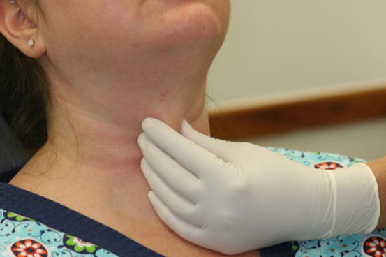 Fig. 1.12Inquire if patient has any difficulty swallowing
 -
Inspect for enlargement or mobility. Listen for hoarseness.
Submandibular neck nodes
-
To palpate the submandibular and submental nodes have the patient lower the chin and manually palpate directly underneath the chin and the medial side of the mandible (Fig.1.13).

