The desire for presurgical management of the cleft lip and palate (CLP) deformity can be traced back several centuries. The goal was to presurgically mitigate the severity of the deformity in hopes of achieving a superior surgical outcome. Most attempts at presurgical management only addressed the alveolar deformity. Until nasoalveolar molding (NAM) was introduced, the nasal deformity that was associated with cleft lip and palate was addressed entirely by surgical means.
This chapter will present the role of NAM in optimizing the primary surgical repair of the nose, lip, and alveolus in infants born with unilateral cleft lip and palate (UCLP) and bilateral cleft lip and palate (BCLP). This chapter will primarily apply to nonsyndromic cleft lip and palate, although NAM can be used to treat certain syndromic cleft cases. Although the principles of treatments are similar for patients with UCLP and BCLP, there are also unique features to each group that deserve specific attention.
Etiopathogenesis/Causative Factors
The cleft lip results from failure of the medial nasal and maxillary processes to fuse between the 4th to the 6th week of fetal development. Clefting of the palate results when the left and right palatal processes fail to elevate and fuse in the midline between the 8th to the 12th week of fetal development.
It is generally accepted that the majority of clefts are a product of both a genetic susceptibility and an environmental insult to the developing fetus. Progress has been made in the genetic determination of clefting and regarding issues related to prenatal care.
Pathologic Anatomy
Clefting of the lip and palate can occur in combination, as an isolated deformity, or as part of a syndrome, and can affect either one (unilateral) or both sides (bilateral) of the face. The severity of each cleft is variable as well and can be either a complete cleft or an incomplete cleft. The classic descriptions below are offered for complete UCLP and BCLP.
Unilateral Cleft Lip and Palate
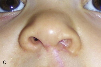
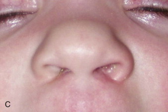
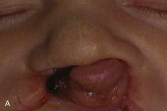
In UCLP, the alveolus is asymmetrically divided into two pieces: the greater alveolar segment (including the premaxilla) and the lesser alveolar segment (on the cleft side). With the greater alveolar segment protruded anteriorly and deviated to the noncleft side, the medial and lateral insertions of the cleft side nasal alar cartilage and the lip segments are drawn apart, resulting in the classic UCLP deformities. Perhaps one of the most disfiguring components of the UCLP deformity (and most challenging to repair) is the prolapsed cleft side alar cartilage, which is often under tension from the malpositioned medial and lateral points of insertion ( Fig. 84-1 ).


Bilateral Cleft Lip and Palate
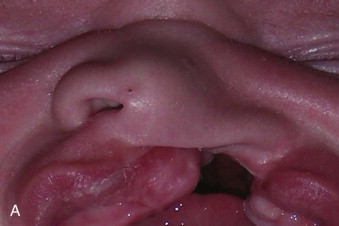
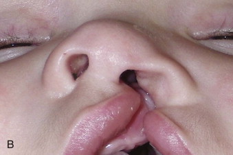
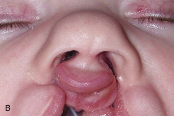
In BCLP, the alveolar ridge is divided into three segments. The premaxillary segment is often positioned outside the mouth. There are two lateral (or posterior) alveolar segments located intraorally. The lip is also divided into three parts. The prolabium appears to originate from the tip of the nose, because the columella is severely deficient. The lateral lip segments are located significantly behind the anteriorly protruding premaxilla and prolabium. This results in a widened nasal tip and nasal base, as the lateral points of insertion of the alar wings are tethered posteriorly with the lips and as the nasal tip is drawn forward with the premaxilla and prolabium ( Fig. 84-2 ).


Pathologic Anatomy
Clefting of the lip and palate can occur in combination, as an isolated deformity, or as part of a syndrome, and can affect either one (unilateral) or both sides (bilateral) of the face. The severity of each cleft is variable as well and can be either a complete cleft or an incomplete cleft. The classic descriptions below are offered for complete UCLP and BCLP.
Unilateral Cleft Lip and Palate
In UCLP, the alveolus is asymmetrically divided into two pieces: the greater alveolar segment (including the premaxilla) and the lesser alveolar segment (on the cleft side). With the greater alveolar segment protruded anteriorly and deviated to the noncleft side, the medial and lateral insertions of the cleft side nasal alar cartilage and the lip segments are drawn apart, resulting in the classic UCLP deformities. Perhaps one of the most disfiguring components of the UCLP deformity (and most challenging to repair) is the prolapsed cleft side alar cartilage, which is often under tension from the malpositioned medial and lateral points of insertion ( Fig. 84-1 ).

Bilateral Cleft Lip and Palate
In BCLP, the alveolar ridge is divided into three segments. The premaxillary segment is often positioned outside the mouth. There are two lateral (or posterior) alveolar segments located intraorally. The lip is also divided into three parts. The prolabium appears to originate from the tip of the nose, because the columella is severely deficient. The lateral lip segments are located significantly behind the anteriorly protruding premaxilla and prolabium. This results in a widened nasal tip and nasal base, as the lateral points of insertion of the alar wings are tethered posteriorly with the lips and as the nasal tip is drawn forward with the premaxilla and prolabium ( Fig. 84-2 ).

Diagnostic Studies
Ultrasonography
At the 20-week structural ultrasonogram that most women have, it is possible to observe most clefts of the lip and palate.
Direct Examination of Cleft Type
Ultimately, it is the surgeon’s decision to include NAM in the treatment protocol if he or she feels a superior primary outcome could be achieved following its use. Then it is up to the orthodontist (or whoever is performing the NAM) to determine if the presurgical result desired by the surgeon can be achieved. At this point, NAM is presented to the family as a treatment alternative and discussed in detail as described later.
Clefting of the nose, lip, alveolus, and palate can have variable presentation. While NAM is appropriate for most cleft types, its use is not indicated in all of them. For example, isolated palatal clefts do not benefit from NAM, because the alveolar ridge is intact and nasal morphology is not affected. On the other hand, partial lip clefts with intact or notched alveolar ridges but with significant nasal deformity can benefit from NAM.
Treatment/Reconstructive Goals
The rationale for performing NAM is to reduce the severity of the initial cleft deformity prior to the primary surgery. In this way, the optimal outcome may be achieved, leading to (1) normalized social interaction at the earliest stage of development possible by reducing the stigmata of living with a cleft, (2) reduction in the need for costly surgical revisions, and (3) a decrease in the cost and burden of cleft rehabilitation on the patient, family, and society.
Specifically, in UCLP, the treatment goals are to (1) reduce the alveolar gap, (2) bring the lip segments into passive contact, (3) increase the surface area of the nasal mucosal lining, (4) reduce nasal septum deviation, and (5) to achieve symmetry between the lower lateral alar cartilages.
Stay updated, free dental videos. Join our Telegram channel
VIDEdental - Online dental courses


