Material and methods
This retrospective study was approved by the institutional review board at the University of Washington. The consecutive sample of orthodontic records was selected from the retention files of the Department of Orthodontics, spanning the years 1995 to 2011. For patients from January 1995 to December 2008, it was possible to identify potential subjects by searching the retention files in the graduate clinic. The retention files provide a summary of each patient’s diagnosis, treatment plan, and final outcomes. After 2008, the retention files were not systematically collected. Therefore, initial models of all consecutively finished patients from 2008 to 2011 were evaluated to identify potential subjects.
The inclusion criteria were defined as patients treated at the orthodontic graduate clinic with a Class II subdivision malocclusion whose initial and final records included complete chart notes, intraoral and extraoral photographs, study models, and cephalometric radiographs. Patients with syndromes or cleft lip and palate were excluded. Categorization as a Class II subdivision required at least a half-cusp difference between the right and left sides. For example, a full-cusp Class II relationship on the right side and a half-cusp Class II relationship on the left side would qualify.
After selecting the eligible subjects, we masked all identifiers on the study casts. Initial maxillary midline position was determined by evaluating patient photographs, and then this evaluation was cross-referenced with diagnostic notes in the chart. Mandibular midlines were related to the maxillary and facial midlines by assessing photographs and study casts. Again, these evaluations were cross-referenced with chart notes. A determination on mandibular skeletal asymmetry was also performed using facial photographs and chart notes. For a few patients, posteroanterior cephalometric radiographs were also available. Final midline assessments were obtained in the same fashion, although chart notes were often not available for cross-referencing. Dental midlines that were within 1 mm to either side of the facial midline were considered to be symmetric with the face.
In a random and blinded fashion, 2 calibrated examiners (S.E.C., S.R.J.) independently scored all initial study casts for the peer assessment rating (PAR) and, starting 1 week later, measured the final casts. PAR scores used in data analysis represented an average of each examiner’s score. For PAR scores for which the examiners disagreed by more than 5 points, each examiner measured the casts again. If the scores still differed by more than 5 points, the examiners met, and a consensus score was obtained.
Periodontal probes were used to measure the initial and final overjet and overbite on each set of casts using the American Board of Orthodontics measuring standards. Digital calipers were used to measure the horizontal millimetric difference in molar relationship from Class I. The measurement was from the maxillary first molar mesiobuccal cusp tip to the mandibular first molar buccal groove. A perfect Class I molar relationship was defined as having a 0-mm discrepancy. Class II molar relationships were denoted as a positive discrepancy, and Class III molar relationships were denoted as a negative discrepancy. To evaluate the success of the molar correction, a Class I or a Class II target molar occlusion was established for each Class II side, based on extractions or missing teeth on the Class II side. For example, a Class II side with no missing teeth and no extractions would have a Class I relationship as the target. A Class I molar target occlusion was assigned a value of 0 mm (distance between the mesiobuccal cusp tip of the maxillary first molar and the buccal groove of the mandibular first molar in a Class I relationship). A Class II molar target was assigned a value of 6.5 mm, using the same measurement landmarks (this value was chosen based on the distribution of patients with a Class II target with respect to the average distance that Class II molars were typically displaced, which was 6.5 mm). Molar targets were considered acceptable if they were within 1 mm of the established target values (0 mm for Class I, and 6.5 mm for Class II).
Initial and final cephalometric radiographs were hand traced, and the following measurements were obtained: maxillary central incisors to NA in millimeters and degrees, mandibular central incisors to NB in millimeters and degrees, and mandibular central incisors to the mandibular plane. All PAR, cephalometric, and midline measurements were performed before the examiner (S.E.C.) knew the patient’s treatment. Each chart was thoroughly examined to abstract information on the research parameters: sex, date, age at bond and debond, and all treatments rendered. Details of the treatments included extractions, headgear, fixed functional appliances, elastics, other auxiliary appliances, and surgery recommended or completed.
Statistical analysis
Means and proportions were calculated for the parameters of interest. Means were compared using analysis of variance and t tests. Proportions were compared using the chi-square test or the Fisher exact test. Results were regarded as significant at P <0.05.
The intraclass correlation coefficient was used to determine interrater reliability for the PAR measurements. Based on 20 randomly selected pairs of measurements, the intraclass correlation coefficient was 0.781, indicating acceptable interrater reliability. For key outcomes, 10 subjects were selected randomly for remeasurement at least 1 month later. Dahlberg’s formula was used to calculate intrarater reliability for midline assessment, mandibular incisor to mandibular plane angulation, and molar class measurement.
The intrarater reliability values for midline deviations, mandibular incisor to mandibular plane angulation, and molar correction were 0.19 mm, 1.23°, and 0.06 mm, respectively.
Results
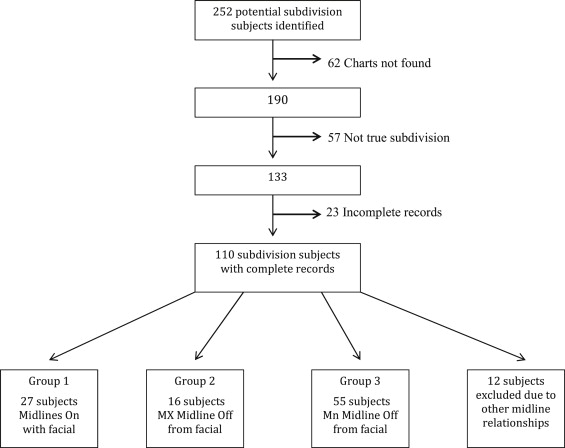
From 1995 to 2008, 218 potential subjects were identified from the retention files ( Fig 1 ). Another 34 potential subjects were identified from assessing models from 2008 to 2011. Sixty-two patients were excluded because their charts could not be located. Another 57 subjects were excluded for failure to meet the subdivision inclusion criterion. Another 23 subjects were lost because of incomplete records. This left a total of 110 subjects, or 44% of those initially identified for potential inclusion.

The subjects were then placed into groups based on similarities in their midline relationships and the etiologies of their asymmetries. Group 1 was composed of subjects whose maxillary and mandibular midlines were coincident with the facial midline; 27 subjects met this criterion ( Fig 1 ). The etiology of the asymmetry in Group 1 was determined to be primarily dental in origin. Group 2 was composed of subjects whose maxillary midlines deviated to 1 side of the facial midline by more than 1 mm, but whose mandibular midlines were coincident with the facial midline; 16 subjects fell into this category, and their etiology was also determined to be primarily dental in origin. Group 3 was composed of subjects whose maxillary midlines were coincident with the facial midlines, but whose mandibular midlines were more than 1 mm from the facial midline; 55 subjects fell into this category, and most were judged to have some degree of skeletal mandibular asymmetry based on chin point deviation. However, 9 subjects in Group 3 had posterior crossbites and potential lateral shifts. Even though all subjects had Class II subdivision molar relationships, they had the following midline relationships.
Group 1: maxillary and mandibular midlines corresponding with the facial midline
Group 2: maxillary midline not corresponding with the facial midline
Group 3: mandibular midline not corresponding with the facial midline
An additional 12 subjects fell into other midline categories, such as both midlines deviated to the same or the opposite side. They were excluded from further analysis because of their small numbers.
At baseline, the 3 main groups had no significant differences in sex distribution, age at banding or bonding, incisor proclination, PAR score, or overbite ( Table I ). Not surprisingly, the 2 groups that were created based on noncoincident midlines exhibited significant differences for their midline measurements ( Table I ). Group 3 also had about 1 mm of additional overjet at the start of treatment.
| Variables | Total sample (n = 98) | Group 1 (n = 27) | Group 2 (n = 16) | Group 3 (n = 55) | P value | ||||
|---|---|---|---|---|---|---|---|---|---|
| Female sex, n (%) | 53 (54) | 16 (59) | 10 (63) | 28 (51) | 0.70 | ||||
| Initial age (y-m), median (mean) | 13-2 (18-9) | 13-9 (17-3) | 15-3 (20-8) | 14-11 (19-7) | 0.57 | ||||
| Mean | SD | Mean | SD | Mean | SD | Mean | SD | ||
| Maxilla to Mx incisors | |||||||||
| Mx1-NA (°) | 20.6 | 8.1 | 18.8 | 7.9 | 21.2 | 7.9 | 21.3 | 8.7 | 0.39 |
| Mx1-NA (mm) | 5.3 | 2.6 | 5.7 | 2.8 | 4.3 | 2.3 | 5.3 | 2.5 | 0.069 |
| Mandible to Mn incisors | |||||||||
| Mn1-NB (°) | 25.9 | 7.6 | 26.6 | 5.2 | 28.4 | 7.7 | 24.8 | 8.8 | 0.27 |
| Mn1-NB (mm) | 6.0 | 2.8 | 5.8 | 2.4 | 7.0 | 2.7 | 5.8 | 2.9 | 0.38 |
| Mn1-MP (°) | 96.2 | 8.0 | 95.9 | 6.1 | 98.1 | 8.1 | 95.9 | 8.7 | 0.75 |
| Mx midline deviation (from facial midline) (mm) | 0.6 | 0.9 | 0.2 | 1.0 | 2.3 | 1.0 | 0.3 | 1.0 | <0.001 |
| Mn midline deviation (from facial midline) (mm) | 1.4 | 1.1 | 0.6 | 1.1 | 0.4 | 1.1 | 2.3 | 1.1 | <0.001 |
| Class II side (mm from Class I) ∗ | 3.6 | 1.3 | 3.4 | 1.3 | 3.7 | 1.2 | 3.6 | 1.3 | 0.76 |
| Class I side (mm from Class I) ∗ | 0.2 | 0.9 | 0.2 | 1.1 | 0.1 | 0.5 | 0.0 | 0.9 | 0.67 |
| Initial overjet (mm) | 5.2 | 2.2 | 4.3 | 2.1 | 4.8 | 2.3 | 5.7 | 2.2 | 0.016 |
| Initial overbite (mm) | 4.6 | 1.9 | 4.4 | 2.2 | 3.9 | 2.2 | 4.9 | 1.7 | 0.10 |
| Initial PAR score | 28.1 | 10.4 | 25.2 | 10.0 | 28.7 | 10.0 | 29.3 | 9.0 | 0.26 |
| Extraction rate | 41% | 48% | 38% | 35% | 0.74 | ||||
Stay updated, free dental videos. Join our Telegram channel

VIDEdental - Online dental courses


