Introduction
This study was performed to investigate the cephalometric configuration of the occlusal plane in patients with anterior open bite.
Methods
Of 61 subjects with open bite (overbite ≥3.75 mm) who had been recruited consecutively from January 2006 to November 2013 and had no history of orthodontic treatment, 14 cephalometric landmarks indicating the incisal edge or the buccal or mesiobuccal cusp tips of each tooth were used for K-means clustering to classify the occlusal plane configuration. For the open-bite group and a control group with normal occlusion (n = 38), dentoalveolar height, which is the perpendicular distance of each tooth to the palatal or mandibular plane, was compared among the clusters and between the 2 groups.
Results
The open-bite subjects were divided into 2 clusters according to occlusal contact of the premolars: Y-form and V-form (with and without premolar contact, respectively). The normalized dentoalveolar heights of the 4 mandibular teeth (lateral incisor to second premolar) were significantly greater in the Y-form class than in the V-form class. The dentoalveolar heights of the 5 maxillary teeth (lateral incisor to first molar) were significantly greater in the open-bite group than in the control group.
Conclusions
For anterior open-bite treatment, the cephalometric configuration of the occlusal plane should be considered based on the occlusal contacts of the premolars.
Highlights
- •
Dentoalveolar tooth heights were compared in open-bite and normal occlusions.
- •
K-clustering was used to divide open-bite subjects into 2 groups, V-form and Y-form.
- •
Dentoalveolar heights of 4 mandibular teeth were greater in the Y-form group.
- •
Dentoalveolar heights of 5 maxillary teeth were greater in the open-bite group.
- •
Open bite may be caused by overeruption of the maxillary molars.
The treatment of anterior open bite (AOB) is challenging because of its high relapse rate. Although surgical and nonsurgical approaches have been proposed for adults, no consensus for the optimal treatment has been established yet. The treatment protocol should be selected in consideration of many factors, including skeletal and dentoalveolar features, esthetics, and patient’s expectations.
For a better understanding of AOB, several authors have investigated its cephalometric features and agreed on some skeletal characteristics such as a decrease in the ratio of posterior to anterior facial height and an increase in lower anterior facial height, Y-axis, gonial angle, and mandibular plane angle. However, there have been some controversies regarding dentoalveolar characteristics such as the interincisal angle, dental heights, and occlusal plane angle. In particular, the occlusal plane angle was generally greater in the AOB group than in the normal occlusion group, but its definition was inconsistent in the studies. Tsang et al addressed 3 occlusal planes: the functional occlusal plane, drawn along the maximum intercuspation of the posterior teeth; the maxillary occlusal plane (MxOP); and the mandibular occlusal plane (MnOP). Taibah and Feteih used the MxOP and the MnOP only, whereas other authors did not clarify the definition of the occlusal plane.
Some researchers have reported that the MxOP and MnOP should be considered separately in AOB patients because the measurement of the occlusal plane angle, by bisecting the AOB, may lead to the wrong conclusion that the AOB produces an increase in the occlusal plane angle. Hence, the MxOP and the MnOP angles were measured separately, as a line connecting the incisal edge of the central incisor and the mesiobuccal cusp tip of the first molar. However, the positions of other teeth composing the occlusal plane have been rarely considered, in spite of their clinical importance. The configuration of each occlusal plane can influence the treatment plan and posttreatment stability. For example, an AOB with 2 distinct occlusal planes can be corrected by orthognathic surgery with routine presurgical orthodontic treatment, segmental surgery, or intrusion of the occluding teeth, and each treatment modality has a different posttreatment stability.
Previous studies have tried to distinguish skeletal open bite from dental open bite on a lateral cephalogram to understand the etiologic factors of this condition. A study suggested that a dental open bite occurs most often in children, and a skeletal open bite in adults ; another study reported that the 2 types of open bite could be distinguished by the growth pattern of the jaw. However, it has been difficult to make a clear distinction between the 2 types. Therefore, it would be better for clinicians to identify the cephalometric configurations rather than the etiologic factors of open bite.
Because the treatment of AOB aims to change the MxOP and the MnOP, a better understanding of the cephalometric configuration of the occlusal plane is essential for correct diagnosis, proper treatment planning, and successful stability. However, there have been only a few studies on the configuration of the occlusal plane in patients with AOB. Therefore, this study was performed to investigate the cephalometric configuration of the occlusal plane in patients with AOB, to measure the height of each tooth from the palatal or mandibular plane, and to compare these heights with those of subjects with normal occlusion.
Material and methods
This study included an open-bite group (n = 61; 16 men, 45 women; mean age, 25.4 years; range, 20.5-42.0 years) and a control group (n = 38; 12 men, 26 women; mean age, 25.5 years; range, 20.4-37.8 years) ( Table I ). The 61 subjects in the open-bite group were selected from a total of 164 orthodontic patients who had visited the Gangnam Severance Dental Hospital in Seoul, Korea, between January 2006 and November 2013, had lateral cephalograms and impressions taken of both arches for the fabrication of dental casts, and had been diagnosed with AOB. The inclusion criteria were moderate-to-severe AOB (≥3.75 mm), no missing teeth except for the third molars, age over 20 years, and mild facial asymmetry with less than 1 mm of occlusal plane canting and less than 2 mm of menton deviation. The exclusion criteria were history of orthodontic treatment, systemic disease, cleft lip or palate, and craniofacial syndromes. The open-bite group included patients with skeletal Class I (n = 6), Class II (n = 52), and Class III (n = 3) malocclusions. The control group was carefully selected from the archives of the orthodontic department of the same hospital with Class I skeletal and dental relationships, a normodivergent facial profile, and less than 2 mm of arch length-tooth size discrepancy. The same inclusion criteria of the open-bite group were applied to the control group, including additionally 2 to 4 mm of overjet and overbite. This study was approved by the institutional review board of the Gangnam Severance Dental Hospital (3-2012-0116).
| Open bite (n = 61) | Control (n = 38) | P value | |
|---|---|---|---|
| Age (y) | 25.36 ± 3.27 | 25.50 ± 6.04 | 0.922 |
| Overbite ∗ (mm) | −5.40 ± 1.56 | 2.80 ± 0.70 | 0.000 † |
| Overjet (mm) | 4.38 ± 2.85 | 3.44 ± 0.39 | 0.278 |
| SNA (°) | 81.08 ± 3.19 | 81.21 ± 2.59 | 0.833 |
| SNB ∗ (°) | 75.04 ± 6.27 | 78.61 ± 2.43 | 0.000 † |
| ANB (°) | 5.42 ± 2.48 | 2.60 ± 0.94 | 0.000 † |
| APDI (°) | 78.49 ± 6.01 | 85.10 ± 3.58 | 0.000 † |
| ODI ∗ (°) | 66.53 ± 7.38 | 71.37 ± 3.71 | 0.000 † |
| Björk sum (°) | 405.70 ± 5.96 | 395.71 ± 2.67 | 0.000 † |
| Gonial angle (°) | 126.95 ± 7.46 | 121.14 ± 4.73 | 0.000 † |
| SN-MP (°) | 45.70 ± 5.96 | 35.71 ± 2.67 | 0.000 † |
| PP-MP (°) | 35.64 ± 5.99 | 25.52 ± 3.54 | 0.000 † |
| PFH/AFH | 58.42 ± 4.55 | 65.23 ± 2.56 | 0.000 † |
| U1-SN (°) | 106.98 ± 7.14 | 106.79 ± 6.97 | 0.895 |
| IMPA (°) | 93.19 ± 7.66 | 94.08 ± 5.93 | 0.542 |
∗ Mann-Whitney U test was used because overbite, SNB, and ODI were not normally distributed. Other variables were analyzed with an independent t test.
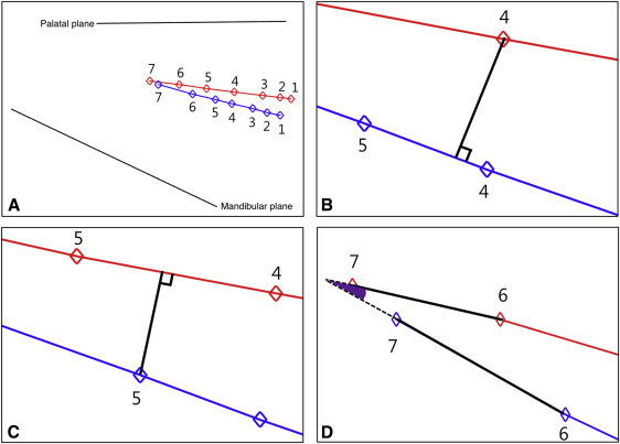
Based on the lateral cephalograms, 19 landmarks were identified and digitized using an image-measuring program (Image-Pro PLUS, version 3.0; Media Cybernetics, Bethesda, Md). Fourteen landmarks indicated the incisal edges or the buccal and mesiobuccal cusp tips of each tooth, and 4 landmarks (anterior nasal spine, posterior nasal spine, lower gonion, and menton) composed 2 reference planes: the palatal plane, which connects the anterior and posterior nasal spines, and the mandibular plane, which connects lower gonion and menton ( Fig 1 , A ). Nasion was also digitized to measure the anterior facial height that was used to normalize distances related to the teeth. Each landmark was coordinated, and linear and angular measurements were performed using MATLAB software (MathWorks, Natick, Mass).

Pattern recognition is a method that automatically detects regularities from given data. K-means clustering is one of such methods that split data into several groups based on their similarities. To measure the similarities, we defined features representing linear and angular relationships of the teeth. Here, 20 features (F 1 –F 20 ) were selected that could be categorized into 3 types: the perpendicular distance from a given maxillary tooth to a corresponding line constructed by 2 adjacent mandibular teeth (F 1 –F 7 ) ( Fig 1 , B ); the perpendicular distance from a given mandibular tooth to a corresponding line constructed by 2 adjacent maxillary teeth (F 8 –F 14 ) ( Fig 1 , C ); and the angle between each maxillary tooth line and the corresponding mandibular tooth line (F 15 –F 20 ) ( Fig 1 , D ). The subscript number of each feature increases from the central incisor to the second molar. Figure 1 shows the position of each tooth in the basal bone and examples of 3 descriptor types. Features F 1 -F 14 were normalized by the anterior facial height of each subject to compensate for their vertical differences and conduct fair comparisons.
Not all features are useful in cluster analysis. Therefore, feature selection is a crucial step in the analysis. In this study, the t test and U test could not be used because the distributions of features were neither normal nor similar to each other. To overcome this difficulty, we used the entropy-based feature ranking method. Entropy is a measure of the average amount of information in each sample drawn from a distribution. For example, if the removal of feature F 1 leads to higher entropy than the removal of feature F 2 , then it is concluded that feature F 1 is more important than feature F 2 . This method uses this removal process in series to select several high-ranking features. Table II shows the ranking of features used in this study. Details on how to determine the ranking are shown in the Appendix.
| Ranking | Variables | Feature | Entropy |
|---|---|---|---|
| 1 | F 12 | Perp distance of MnPM2 | 529.18 |
| 2 | F 5 | Perp distance of MxPM2 | 528.98 |
| 3 | F 4 | Perp distance of MxPM1 | 528.77 |
| 4 | F 11 | Perp distance of MnPM1 | 528.71 |
| 5 | F 13 | Perp distance of MnM1 | 528.64 |
| 6 | F 6 | Perp distance of MxM1 | 528.39 |
| 7 | F 20 | Angle between a line connecting MxM1 and MxM2 and a line connecting MnM1 and MnM2 | 528.17 |
| 8 | F 14 | Perp distance of MnM2 | 528.00 |
| 9 | F 10 | Perp distance of MnC | 527.90 |
| 10 | F 9 | Perp distance of MnI2 | 527.78 |
| 11 | F 7 | Perp distance of MxM2 | 527.75 |
| 12 | F 8 | Perp distance of MnI1 | 527.66 |
| 13 | F 3 | Perp distance of MxC | 527.59 |
| 14 | F 1 | Perp distance of MxI1 | 527.56 |
| 15 | F 2 | Perp distance of MxI2 | 527.48 |
| 16 | F 18 | Angle between a line connecting MxPM1 and MxPM2 and a line connecting MnPM1 and MnPM2 | 526.94 |
| 17 | F 16 | Angle between a line connecting MxI2 and MxC and a line connecting MnI2 and MnC | 526.55 |
| 18 | F 19 | Angle between a line connecting MxPM2 and MxM1 and a line connecting MnPM2 and MnM1 | 526.34 |
| 19 | F 17 | Angle between a line connecting MxC and MxPM1 and a line connecting MnC and MnPM1 | 526.23 |
| 20 | F 15 | Angle between a line connecting MxI1 and MxI2 and a line connecting MnI1 and MnI2 | 525.98 |
To determine the optimal number of features and clusters for K-means clustering, we used 2 criteria: the Calinski-Harabasz criterion and the mean of silhouette values. A higher Calinski-Harabasz index indicates better-defined clusters, and the silhouette value shows how well the data lie within the clusters. We added high-ranking features one by one and checked the above criteria to determine the optimal number of features.
Based on the number of clusters and high-ranking features, an expert (Y.J.C.) with more than 10 years of experience classified the open-bite subjects using dental casts, and the results were compared with those from the cluster analysis.
The dentoalveolar heights of each tooth, which were the perpendicular distances to the palatal and mandibular planes in the maxilla and the mandible, respectively, were calculated and compared between the open-bite and control groups using the Mann-Whitney U test. Based on the cluster analysis, the comparisons among subgroups in each cluster were also performed with the same test.
A random selection of 10 lateral cephalograms from each group was digitized twice within 2 weeks by 1 examiner (Y.J.C.), and intraclass correlations were calculated. The correlations ranged from 97.3% to 99.5%; hence, the first reference points were used.
Results
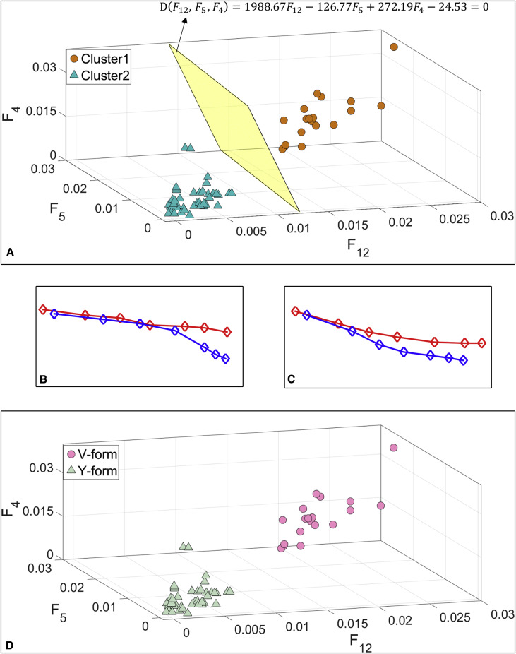
The open-bite subjects were divided into 2 clusters using the top 3 features ( Table III ). Figure 2 , A , shows the Calinski-Harabasz index values for the different clusters using the top 3 features, and Figure 2 , B , shows the corresponding silhouette plot of the 2 clusters. The top 3 features were F 12 , F 5 , and F 4 , which indicated the perpendicular distances of the mandibular second premolar, the maxillary second premolar, and the maxillary first premolar to a corresponding line constructed by 2 adjacent teeth in the opposite dentition, respectively ( Table II ).
| Number of most important features | Optimum number of cluster (C-H criterion) | Quality of cluster analysis (mean of silhouette values) |
|---|---|---|
| 1 | 10 | 0.7515 |
| 2 | 10 | 0.6542 |
| 3 ∗ | 2 ∗ | 0.8653 ∗ |
| 4 | 2 | 0.8409 |
| 5 | 2 | 0.8262 |
| 6 | 2 | 0.8149 |
| 7 | 10 | 0.7900 |
| 8 | 10 | 0.7900 |
| 9 | 10 | 0.7897 |
| 10 | 10 | 0.7895 |
| 11 | 10 | 0.7895 |
| 12 | 9 | 0.7997 |
| 13 | 10 | 0.7890 |
| 14 | 10 | 0.7887 |
| 15 | 10 | 0.7885 |
| 16 | 10 | 0.6190 |
| 17 | 10 | 0.5878 |
| 18 | 2 | 0.6021 |
| 19 | 3 | 0.4584 |
| 20 | 2 | 0.3692 |

Figure 3 , A , demonstrates the division of the 2 clusters based on the top 3 features, and Table IV shows the values of these features in each cluster. All values in cluster 1 were much higher than those in cluster 2, which were close to zero. This indicated that in cluster 1 the maxillary first and second premolars and the mandibular second premolar were out of occlusion; in cluster 2, the teeth had occlusal contacts with the opposite teeth. Figure 3 , B and C , illustrates typical figures of each cluster. Based on these figures, we named clusters 1 and 2 the Y-form class and the V-form class, respectively. Figure 4 , A and B , shows typical intraoral photographs of the Y-form and V-form classes, respectively, and Figure 4 , C , depicts the top 3 features.


