
Management of cleidocranial d ysplasia I—t reatment of an a dolescent patient
CASE STORY
A 17-year-old male patient presents with a chief complaint of “I don’t like the way my teeth look, and I would like to have a nice smile.” The patient is accompanied by his mother (see Case 37) and has been diagnosed with cleidocranial dysplasia (CCD). The patient has received only palliative and emergency care for his dental condition and no formal management plan has been pursued for the treatment of CCD.
Figures 1, 2, and 3: intercuspation.
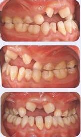
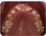
Figure 4: Maxillary occlusal view.
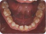
Figure 5: Mandibular occlusal view.
LEARNING GOALS AND OBJECTIVES
- To recognize signs of CCD
- To discuss treatment approaches for a young patient with CCD
- To understand the advantages and disadvantages of various treatment options
- To manage CCD dental treatment with a multidisciplinary approach
Medical History
- Cleidocranial dysplasia
Dental History
- Palliative treatment including extraction of mobile primary teeth
Medications and Allergies
- No medications
- No known drug allergies
Review of Systems
- Vital signs:
- Blood pressure: 120/78
- Heart rate: 70 beats/minute
- Respiration rate: 16 breaths/minute
Significant Soft Tissue Examination Findings
- No significant findings
Significant Clinical Findings/Problem List
- Anterior open bite
- Bilateral posterior cross-bite
- Underdeveloped maxilla, class III malocclusion
- Retention of primary teeth
- Supernumerary teeth
- Poor anterior aesthetics
- Plaque accumulation
- Deficiency in speech
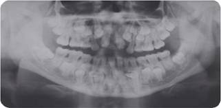
Radiographic Findings
Figure 6: Panoramic radiograph.
Charting
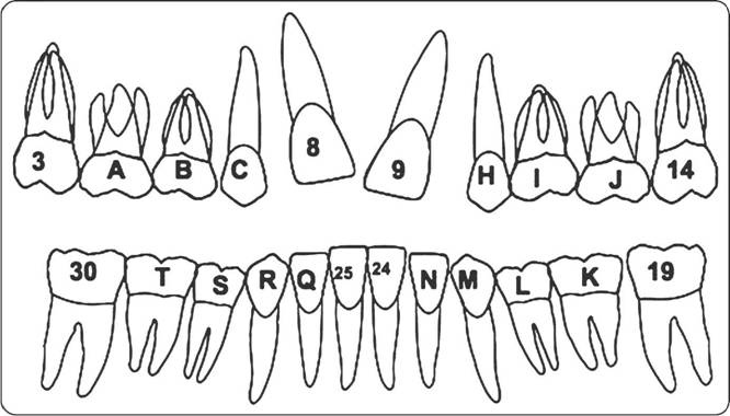
(See Fig. 7.)
Diagnosis
- Cleidocranial dysplasia
- Deficient aesthetics
Figure 7: Dental charting of erupted teeth.
Clinical Decision-Making Determining Factors
- Cleidocranial dysplasia (CCD) is an autosomal dominant condition that is genetically inherited. The patient presented here has inherited this condition from his mother (see Case 37). The genetic condition affects bone growth, often causing aplasia (defective development) or hypoplasia (incomplete or underdevelopment) of bones undergoing intramembranous ossification. The general appearance of a person presenting with CCD is typically frontal, parietal, and occipital bossing; underdevelopment of the maxilla; and a brachycephalic appearance of the skull. The clavicles are among the bones commonly affected by this condition (Gonzalez Lopez, Ortiz Solalinde et al. 2004; Angle and Rebellato 2005; Bratu 2005),
- Dental findings in patients with CCD may include class III jaw relationship, underdeveloped maxilla, overretention of deciduous teeth, failure or delayed eruption of permanent teeth, multiple supernumerary teeth, and cyst formation. Because dental complications have a direct effect on the patient’s quality of life, CCD patients are often in extensive dental treatment that includes multiple disciplines (Golan, Baumert et al. 2004; Angle />
Stay updated, free dental videos. Join our Telegram channel
VIDEdental - Online dental courses


