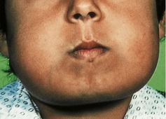
Case• 63. A child with a swollen face
SUMMARY
A 5-year-old boy has painless bilateral facial swellings. Identify the cause and recommend treatment.
History
Complaint
The patient is brought by his parents who have noticed that his face has become fat. They are concerned about his appearance and say that he is being teased and bullied at school.
History of complaint
His parents say that the patient has had a chubby face since he was a toddler but that the swelling has become more noticeable over the last 2 years. He is in no pain.
Medical history
He is otherwise fit and well, has had all recommended immunizations and amongst the childhood illnesses has suffered only chicken pox. His medical practitioner has given him a general examination and found no systemic illness but has referred him to you for a further opinion.
Examination
Extraoral examination
The appearance of the child is shown in Figure 63.1. He appears healthy but has obvious bilateral enlargement of the side of the face. The temporomandibular joints appear normal on palpation. Some upper deep cervical lymph nodes are palpable bilaterally. They are only slightly enlarged, not tender and are freely mobile.
▪ On the basis of what you know, what types of lesion would you consider?
From this view alone it is difficult to tell whether the swelling originates in the salivary glands, mandible or soft tissues. Each site would have different possible causes:
| Condition | Possible causes |
|---|---|
| Soft tissue enlargement | Masseteric hypertrophy is possible. Bruxism is common in children though significant masseteric hypertrophy is rare. |
| Salivary gland enlargement | Rare in children. HIV salivary cystic disease is seen in HIV infection. Mumps can be excluded. Mumps is acute and, in addition, the child would have had mumps vaccine with the rest of the routine childhood vaccinations. |
| Enlargement of the mandible | A few rare inherited disorders of bone could cause bilateral expansion of the ramus. |
| A developmental syndrome | Many syndromes have craniofacial signs and this is a possibility which should be borne in mind. There appear to be no associated features. |
Intraoral examination
Intraoral examination reveals a minimally restored dentition and healthy oral mucosa. Palpation of the mandibular rami shows that they are the source of the enlargement. There is obvious rounded swelling of the posterior body and ramus of the mandible. The lower right second deciduous molar is missing.
Stay updated, free dental videos. Join our Telegram channel

VIDEdental - Online dental courses


