Case• 61. Worn front teeth
SUMMARY
A 60-year-old man presents at your general dental practice saying that his teeth have worn down. What is the cause and how should he be managed?
History
Complaint
The patient is unhappy about his short and discoloured upper anterior teeth. He is also finding that he has difficulty eating. The appearance of his teeth has recently become more important to him because he has taken a job in which he deals with the public. He wishes primarily to improve his appearance and appears to be sufficiently motivated to complete a course of complex dental treatment.
History of complaint
The patient has only recently started to attend his dentist regularly, after a 10-year period without treatment. Most of his posterior teeth were extracted before the age of 45 and he has worn his present upper partial denture for at least 12 years.
Examination
Extraoral examination
No submandibular or cervical lymph nodes are palpable, the temporomandibular joints appear normal and there is no tenderness around the muscles of mastication. Despite the anterior toothwear there is no evidence of loss of occlusal vertical dimension.
Intraoral examination
The oral mucosa is healthy. In the mandible all teeth between the left and right second premolars are present but in the maxilla there are only five anterior teeth remaining. The appearances are shown in Figure 61.1
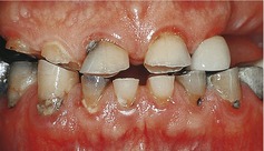
Wear of the incisal edges of upper and lower anterior teeth has produced short clinical crowns and the upper right lateral incisor and canine are worn almost to gingival level. There are deposits of plaque around the cervical margins of his teeth and a number of teeth have cervical caries. Despite the plaque, there is only occasional interdental bleeding on probing and no significant increase in pocket depths. The upper ridges are not extensively resorbed and are broad and well defined. When asked to bite his teeth together the patient adopts the forward mandibular posture shown in Figure 61.1.
Both lower lateral incisors and the lower left canine and first premolar appear discoloured. Only the lower left second premolar fails to respond to an electronic test of vitality.
The patient produces his acrylic upper partial denture from his pocket. It is poorly retentive and of indifferent fit.
▪ What is your differential diagnosis? What features suggest each possibility?
The patient is suffering toothwear. This is usually caused by a combination of three basic underlying processes: erosion, attrition and abrasion. In this case it seems likely that the cause is predominantly erosion with attrition as a secondary factor.
| Aetiological factor | Features |
|---|---|
| Erosion | Erosion is usually caused by excessive dietary acid or regurgitation. Both possibilities must be excluded by careful questioning and dietary analysis (see also Case 60). The appearance of the wear facets suggests erosion as the major cause. Although the teeth interdigitate on incisal enamel, the dentine has been lost from an area which is not in contact with the opposing teeth. |
| Attrition | Attrition is usually caused by occlusal wear and a minor degree is normal. Bruxism and other parafunctional habits may have caused increased attrition. There is no evidence of masticatory muscle tenderness or hypertrophy to suggest that such habits contribute. |
| Abrasion | Abrasion of the teeth is wear by an external agent and is seen when a coarse diet is eaten, as in developing countries, or as a result of toothbrush abrasion.There is nothing to suggest an unusual diet in this case, though the possibility should be excluded by questioning. The pattern is not consistent with a primarily abrasive process. |
Investigations
▪ What investigations would you perform? Why?
Dietary analysis is required to determine whether there is excessive dietary acid and to identify the sources of sugars responsible for the caries in several teeth.
Radiographic assessment by means of either a panoramic tomograph or full mouth periapical films is necessary. In view of the fact that the patient has not attended a dentist for a decade, the panoramic tomograph would provide a useful survey of both teeth and edentulous ridges. Periapical films of all standing teeth are a reasonable alternative in this case because the missing teeth reduce the number of films and radiation dose required. A periapical film of the lower left second premolar is required to assess the feasibility of root canal treatment and obtaining films of the other discoloured teeth would be prudent despite their apparent vitality. Periapical films taken with a paralleling technique would allow accurate assessment of bone levels.
Upper and lower study models are required to assess the treatment options for the short clinical crowns and to design a new denture. When restoring extensively worn teeth the models should be mounted in the retruded contact position on either a fully or semiadjustable articulator. Obtaining an accurate occlusal record is particularly important in this case because the patient has a habi/>
Stay updated, free dental videos. Join our Telegram channel
VIDEdental - Online dental courses


