Case• 59. Difficulty in opening the mouth
SUMMARY
A 40-year-old Indian man presents to you in your general dental practice with limitation of mouth opening. You must identify the cause and institute appropriate follow up.
History
Complaint
The patient complains of difficulty in eating. He cannot open his mouth widely enough to place a proper mouthful of food inside and also has difficulty chewing.
History of complaint
He has noticed the reduction of his mouth opening over a period of several years but has never sought advice. It has not been painful though he has felt a burning sensation from his oral mucosa on eating during the same period. This varies in intensity.
Medical history
The patient is otherwise fit and well.
▪ What are the causes of limitation of mouth opening and how may they be classified?
Limitation of opening is most frequently caused by trismus. By definition, trismus is spasm of the muscles of mastication, though the term is often used loosely when opening is prevented by oedema or inflammation of the muscles or joint. Trismus is usually temporary.
Permanent limitation of opening may be caused by scarring of the soft tissues around the joint or mandible or by fusion of the condyle to the glenoid fossa (ankylosis). The causes may be divided as follows:
| Trismus |
Inflammation in and around the temporomandibular joint
Trauma (fractures and/or soft tissue injury)
Tetanus and tetany
Temporomandibular joint (myofascial) pain dysfunction syndrome
Soft tissue infection around the jaws or joint (usually dental in origin)
|
| Permanent limitation of opening |
A. Extra-articular causes
Fibrosis due to burns or irradiation
Oral submucous fibrosis
Mucosal scarring (e.g. in epidermolysis bullosa)
|
|
B. Intra-articular causes
Congenital ankylosis
Traumatic ankylosis
Ankylosis following pyogenic arthritis
Ankylosis following juvenile arthritis
Neoplasms and other causes of enlargement of the condyle
|
▪ What questions would you ask?
The patient should be asked whether there has been trauma or irradiation to the skull, temporomandibular joint or face and whether there have been any episodes of swelling of the face or around the joint. He should also be asked whether he uses betel quid (pan or paan).
The patient gives no history of trauma, irradiation, inflammation or infection. However, he has been a betel quid chewer for more than 20 years.
Examination
Extraoral examination
The patient looks mildly anaemic. There is no facial asymmetry or evidence of scars or inflammation around the joint. No tenderness can be elicited from the muscles of mastication. There are no clicks or crepitus or tenderness associated with the temporomandibular joint and no mandibular deviation on opening the mouth.
▪ What measurement would you take?
The maximum voluntary mouth opening. The normal interincisal opening in an adult is approximately 30–40 mm. Measurement will provide a baseline reading against which to judge treatment or progression. It will also indicate the feasibility of dental and other intraoral treatment.
This patient can achieve an interincisal opening of 17 mm.
Intraoral examination
▪ The oral mucosa is shown inFigures 59.1and59.2. What do you see?

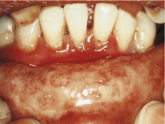 |
| Fig. 59.2 |
The buccal and soft palate mucosae are paler than normal though some mucosal pigmentation consistent with the patient’s skin colour makes this less obvious. When the mouth is opened fully, thin white hard bands run vertically just below the buccal mucosa. These are just visible in the picture and are much more readily felt as hard ridges. Some of the less pale areas are red and atrophic.
Stay updated, free dental videos. Join our Telegram channel
VIDEdental - Online dental courses


