Case• 41. A blister on the cheek
SUMMARY
A 58-year-old lady patient of your general dental practice complains of a sore mouth with blisters. Identify the cause and outline appropriate management.
History
Complaint
The patient complains of a very sore mouth. She describes blisters which last a few hours before bursting to release a clear fluid, or sometimes blood. The palate is particularly affected though lesions may develop anywhere in the mouth and often follow minor trauma. Each blister heals very slowly and the area is painful until healing is complete. She often finds she cannot brush her teeth.
History of complaint
The symptoms started about 1 year ago and are worsening.
Medical history
She has had hypertension for many years and her elderly medical practitioner has been treating her with methyldopa.
Examination
Extraoral examination
A fit-looking woman with a blood pressure of 140/90 when sitting. Visible skin and nails appear normal.
Intraoral examination
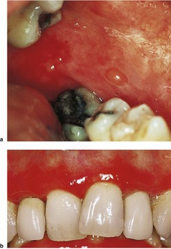
▪ The appearance of the buccal mucosa and gingivae is presented inFigure 41.1. What do you see?
 |
| Fig. 41.1 |
The buccal mucosa has an extensive area of red atrophic mucosa posteriorly, possibly with small ulcers towards the anterior edge. The red area has an irregular margin. A small blister, a few millimetres across, lies near the centre of the buccal mucosa, just above the buccal cusp of the second premolar in the photograph.
The gingivae are also red but no blisters are present. The red area extends from the gingival margin across the mucogingival junction to involve the adjacent alveolar mucosa. The margin is poorly defined. The gingivae around all visible teeth are involved and the distribution of inflammation is not consistent with plaque accumulation as the cause.
Differential diagnosis
▪ Which conditions cause oral blisters?
• Mucous membrane pemphigoid
• Pemphigus vulgaris
• Lichen planus
• Erythema multiforme
• Angina bullosa haemorrhagica
• Epidermolysis bullosa
• Dermatitis herpetiformis
• Viral infections
• Trauma.
A band of red atrophic or eroded mucosa affecting the attached gingiva is known as desquamative gingivitis. Unlike plaque-induced inflammation it is a dusky red colour and extends beyond the marginal gingiva, often to the full width of the attached gingiva and sometimes onto the alveolar mucosa. Some reserve the term for cases where the epithelium blisters or peels while others use it whenever the characteristic red appearance is present.
▪ What are the main causes of desquamative gingivitis?
• Lichen planus
• Mucous membrane pemphigoid
• Pemphigus.
▪ Which of these conditions would you include in your initial differential diagnosis? Explain why.
Either pemphigoid or pemphigus is the most likely diagnosis whenever there is a good history of blister formation. The most frequent cause of oral blisters is mucous membrane (‘cicatricial’) pemphigoid. This typically affects middle-aged and elderly women causing relatively long-lived vesicles and bullae (bullae are blisters greater than 10 mm in diameter) in areas of friction. Pemphigus vulgaris is less common and predominantly affects women in earl/>
Stay updated, free dental videos. Join our Telegram channel
VIDEdental - Online dental courses


