Case• 33. First permanent molars
SUMMARY
A 7-year-old girl has pain from a first permanent molar. What is the cause and how might it affect her dental development?
History
Complaint
The patient’s mother reports that the child suffers intermittent spontaneous discomfort from the upper left teeth.
History of complaint
The symptoms have been vague, no sleep has been lost and there has been no facial swelling. The patient has complained of the pain three or four times over the last month.
Medical history
The child is fit and well.
Dental history
The child has been a regular patient since the age of 3. She has required restorations in four primary molars, one requiring local analgesia. Despite intensive preventive advice and diet analysis, new carious lesions have been present at each recall visit.
Examination
You ask the child to point to the painful tooth and she points to an apparently sound upper left primary canine.
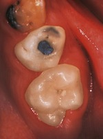
▪ The appearance of the upper left quadrant is shown inFigure 33.1. What do you see?
• An amalgam restoration with ditched or raised margins on the palatal aspect of the first primary molar.
• An apparently sound amalgam restoration in the second primary molar.
• Possible caries in an occlusal pit on the second primary molar.
• An erupting first permanent molar with the occlusal surface not fully through the mucosa.
• A small occlusal cavity in the confluence of the mesial fissures of the permanent molar.
• Plaque or food debris in the fissures.
 |
| Fig. 33.1 |
▪ How do you interpret the information so far and what are the likely diagnoses?
The child is probably pointing at the wrong tooth. The canine appears intact and children are often poor historians. They often have difficulty in localizing the source of pain if the pain is not present at the time of examination.
Pulpitis appears likely because the pain appears poorly localized and is relatively intermittent. A history of hot or cold or sweet exacerbating factors would point to this diagnosis. The likely causes are caries beneath a restoration or carious or traumatic pulpal exposure in one of the primary molars. Any primary molar with an unrestored carious cavity or even a clinically sound restoration should be examined closely for signs of pulpal necrosis.
▪ What features might suggest a necrotic pulp?
• Extension of caries or fracture into the pulp
• Discolouration of the crown
• Swelling or tenderness in the buccal sulcus adjacent to the tooth
• Pus draining from a sinus in the mucosa, usually buccally but occasionally lingually or palatally
• Pus draining from the gingival margin
• Facial swelling
• Well-localized pain
None of these symptoms and signs is present. Pulpitis seems likely.
Investigations
▪ What investigations are indicated? Why?
• Bitewing radiographs to check the proximity of restorations to the pulps, the extent of the occlusal caries in the permanent molar and to detect small proximal surface carious lesions.
• Clinical examination of the other permanent molars for caries.
• Tests of vitality of primary and permanent molars are unlikely to help because the results are unreliable in children.
▪ The left bitewing radiograph is shown inFigure 33.2. What do you see? (Table 33.1)
Stay updated, free dental videos. Join our Telegram channel
VIDEdental - Online dental courses


