Case• 2. A multilocular radiolucency
SUMMARY
A 45-year-old African man presents in the accident and emergency department with an enlarged jaw. You must make a diagnosis and decide on treatment.
History
Complaint
The patient’s main complaint is that his lower back teeth on the right side are loose and that his jaw on the right feels enlarged.
History of complaint
The patient has been aware of the teeth slowly becoming looser over the previous 6 months. They seem to be ‘moving’ and are now at a different height from his front teeth, making eating difficult. He is also concerned that his jaw is enlarged and there seems to be reduced space for his tongue. He has recently had the lower second molar on the right extracted. It was also loose but extraction does not seem to have cured the swelling. Although not in pain, he has finally decided to seek treatment.
Medical history
He is otherwise fit and healthy.
Examination
Extraoral examination
He is a fit-looking man with no obvious facial asymmetry but a slight fullness of the mandible on the right. Palpation reveals a smooth rounded bony hard enlargement on the buccal and lingual aspects. Deep cervical lymph nodes are palpable on the right side. They are only slightly enlarged, soft, not tender and freely mobile.
Intraoral examination
▪ What do you see inFigure 2.1?
 |
| Fig. 2.1 |
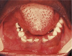
There is a large swelling of the right posterior mandible visible in the buccal sulcus, its anterior margin relatively well defined and level with the first premolar. The lingual aspect is not visible but the tongue appears displaced upwards and medially suggesting significant lingual expansion. The mucosa over the swelling is of normal colour, without evidence of inflammation or infection. There are two relatively small amalgams in the lower right molar and second premolar
If you could examine the patient you would find that all his upper right posterior teeth are extracted and that the lower molar and premolars are 2–3 mm above the height of the occlusal plane. Both teeth are grade 3 mobile but both are vital.
▪ What are the red spots on the patient’s tongue?
Fungiform papillae. They appear more prominent when the tongue is furred, as here, for instance when the diet is not very abrasive.
▪ On the basis of what you know so far, what types of condition would you consider to be present?
The history suggests a relatively slow-growing lesion, which is therefore likely to be benign. While this is not a definitive relationship, there are no specific features suggesting malignancy, such as perforation of the cortex, soft tissue mass, ulceration of the mucosa, numbness of the lip or devitalization of teeth. The character of the lymph node enlargement does not suggest malignancy.
The commonest jaw lesions that cause expansion are the odontogenic cysts. The commonest odontogenic cysts are the radicular (apical inflammatory) cyst, dentigerous cyst and odontogenic keratocyst. If this is a radicular cyst it could have arisen from the first molar, though the occlusal amalgam is relatively small and there seems no reason to suspect that the tooth is nonvital. A residual radicular cyst arising on the extracted second or third molar would be a possibility. A dentigerous cyst could be the cause if the third molar is unerupted. The possibility of an odontogenic keratocyst seems unlikely, because these cysts do not normally cause much expansion. An odontogenic tumour is a possible cause and an ameloblastoma would be the most likely one, because it is the commonest, and arises most frequently at this site and in this age group. There is a higher prevalence in Africans than other racial groups. An ameloblastoma is much more likely than an odontogenic cyst to displace the teeth and make them grossly mobile. A giant cell granuloma and numerous other lesions are possibilities but are all less likely.
Investigations
▪ Radiographs are obviously indicated. Which views would you choose? Why?
| Radiographic view | Reason |
|---|---|
| Panoramic radiograph or an oblique lateral | To show the lesion from the lateral aspect. The oblique lateral would provide the better resolution but might not cover the anterior extent of this large lesion. The panoramic radiograph would provide a useful survey of the rest of the jaws but only that part of this expansile lesion in the line of the arch will be in focus. An oblique lateral view was taken. |
| A posterior-anterior (PA) of the jaws | To show the extent of mediolateral expansion of the posterior body, angle or ramus. |
| A lower true (90°) occlusal | To show the lingual expansion which will not be visible in the PA jaws view because of superimposition of the anterior body of the mandible. |
| A periapical of the lower right second premolar and the first molar | To assess bone support and possible root resorption. |
Several different views are necessary to show the full extent of the lesion. These are listed in the ‘Radiographic view’ table above.
▪ These four different views are shown inFig. 2.2, Fig. 2.3, Fig. 2.4 and Fig. 2.5. Describe the radiographic features of the lesion (shown in ‘Feature of lesion’ table onp. 9).
| Feature of lesion | Radiographic finding |
|---|---|
| Site | Posterior body, angle and ramus of the right mandible. |
| Size | Large, about 10 × 8 cm, extending from the second premolar, back to the angle and involving all of the ramus up to the sigmoid notch, and from the expanded upper border of the alveolar bone down to the inferior dental canal. |
| Shape | Multilo/> |
Stay updated, free dental videos. Join our Telegram channel
VIDEdental - Online dental courses


