Introduction
This study was conducted to determine whether variations in the morphology, size, or shade of maxillary canines would influence perceptions of smile attractiveness in patients with canines substituted for missing maxillary lateral incisors.
Methods
A smiling photograph of a hypodontia patient who had had orthodontic space closure with maxillary canines replacing the lateral incisors was digitally modified to create a bilaterally symmetrical image. Four groups of images were created, digitally altering canine gingival height, crown tip height, canine width, and canine shade. Three groups of judges (40 orthodontists, 40 dentists, and 40 laypeople) ranked the images for smile attractiveness, also scoring the most and the least attractive of each of the 4 groups, and the most and least attractive of all images.
Results
Canine gingival height was the most attractive 0.5 mm below the gingival margin of the maxillary central incisor and progressively less attractive with increasing gingival height. Increasing canine width, increased canine tip height, and pointed canines were perceived to be unattractive. Brighter than normal shades of canines were preferred to darker shades. Narrow canine crowns were most frequently ranked as the most attractive overall, 1.5 mm narrower was preferred by the orthodontists and dentists, and 3.0 mm narrower was preferred by the laypeople. All 3 groups ranked the darkest image, 20 times darker than the original, most frequently as the least attractive image overall. There was good general agreement between orthodontists, dentists, and laypeople for all 4 parameters of smile attractiveness, although laypeople demonstrated greater intragroup variations.
Conclusions
The morphology, size, and shade of the maxillary canine in patients having orthodontic space closure and lateral incisor substitution can have a marked effect on perceived smile attractiveness.
It has been reported that hypodontia (excluding third molars) affects between 3% and 6% of the general population. The maxillary lateral incisor is the second most affected tooth with an incidence of 1% to 2% in Caucasians and accounts for 20% of all missing teeth. The absence of the maxillary lateral incisors affects the proportions of the maxillary labial segment and ultimately the esthetics of the smile. An unattractive smile can affect a person’s appearance, personality, and psychological well-being. It might also provoke an unfavorable response from others in society.
The management of patients with missing maxillary lateral incisors can be challenging, commonly involving 1 of 3 possible treatment approaches: accepting the space, creating enough space for replacement with prosthetic units (denture, bridge, or implant) for the missing lateral incisor, or closing the space orthodontically with the maxillary canine substituting for the missing lateral incisor and camouflaging the canine to mimic the appearance of a lateral incisor. The difficulty with the canine substitution method is achieving an acceptable esthetic outcome, because of the inherent size, shape, and shade differences between the maxillary canine and lateral incisors.
The advantages of space closure with canine substitution are the avoidance of long-term maintenance of the prosthetic replacement of the lateral incisor with a denture, bridge, or implant, which can have future retreatment requirements and cost implications. Patients who had space closure were found to be periodontally more healthy than those with prostheses ; there was no significant difference in occlusal function and prevalence of temporomandibular dysfunction. However, this treatment approach requires careful consideration of the differences in the morphology between the lateral incisor and the canine, and whether the patient is ultimately suitable for space closure and the result will be acceptable and esthetic.
The lateral incisor has incisiform morphology: ie, a smaller, flat-faced tooth compared with the caniniform and conical shaped canine. The canine has a broader neck and, because the tooth is thicker and contains more dentin, is often darker. The gingival margin of the canine is usually higher than that of the lateral incisor, and canines tend to have a prominent tip. If these differences are not compensated for, the esthetic outcome will be compromised.
Several techniques can be used to mask the differences between the 2 tooth types. The canine bracket can be inverted to increase palatal root torque, which might reduce the eminence of the canine. The bracket can be positioned more gingivally to extrude the canine and its gingival margin; the canine tip can then be reduced. Since the canine is wider than the lateral incisor, its width can be reduced mesiodistally with interdental enamel reduction. Finally, because the canine is often naturally darker than the adjacent lateral incisor, the tooth can be bleached after orthodontic treatment.
Previously, authors have investigated perceived smile attractiveness in hypodontia patients ( Table I ). However, the influence of the morphology of the maxillary canine on the perceived attractiveness of the smile has not been investigated.
| Topic | Authors | Sample | Results |
|---|---|---|---|
| Influence of various dimensions of maxillary lateral incisors to smile attractiveness | Bukhary et al | 41 hypodontia and 46 nonhypodontia patients, 30 dentists | Digitally altered width and height of maxillary laterals: 67% then 72% lateral to central ratio preferred. Images of maxillary laterals 1-1.5 mm shorter than the centrals were perceived as attractive; significantly shorter or longer teeth were unattractive. |
| Bilateral aplasia of maxillary lateral incisors and orthodontic space closure | Zimmer and Seifi-Shirvandeh | 25 patients (15 female, 10 male), bilateral maxillary lateral incisor aplasia treated by PPM (for space closure) | Class III elastics are an essential part of PPM that have minor dental and skeletal effects. Orthodontic space closure for bilateral maxillary lateral incisor aplasia by PPM is a valid alternative to prosthetic replacements. |
| Missing maxillary lateral incisors; esthetic impact, comparing dental specialists’ and laypeople’s opinions | Armbruster et al | 140 dentists, 43 orthodontists, 29 dental specialists, 40 laypeople | Laypeople ranked canine substitution highest. Orthodontists preferred no teeth missing >canine substitution >bridges >implants. Restorative dentists would restore the missing tooth, but they did not necessarily rank the restored option image as attractive. |
| Lateral incisor agenesis | Robertson and Mohlin | 50 patients; 30 had space closure, 20 had space opening and restorative replacement | Space closure patients were more satisfied with treatment outcome than space opening patients. No difference in TMD symptoms. Space opening: poorer periodontal condition, greater plaque accumulation, and gingivitis. |
The principal aims of this study were to (1) quantitatively score smile attractiveness in a hypodontia patient, managed with space closure and maxillary canines substituted for the missing lateral incisors, with incremental alteration of the following parameters of the maxillary canine: crown width, crown height and tip morphology, gingival margin height, and shade; and (2) compare the perceptions of smile attractiveness between orthodontists, dentists, and laypeople.
Material and methods
One hundred twenty observers were recruited as participants for the study. They were orthodontists, dentists, and laypeople.
Each group had 40 observers. The orthodontists comprised final year and higher orthodontic trainees, specialist practitioners, and hospital consultants and included 12 men and 28 women (18 white and 22 nonwhite; mean age, 33.9 ± 7.8 years; range, 26-65 years). The dentists were all qualified practitioners and included 16 men and 24 women (26 white and 14 nonwhite; mean age, 34.3 ± 11.2 years; range, 23-57 years). The lay group comprised people from various nonclinical backgrounds and included 11 men and 29 women (33 white and 7 nonwhite; mean age, 36.6 ± 11.7 years; range, 20-63 years). Each observer was asked to rank a series of images in the order of attractiveness of the smiling mouth, displaying the maxillary second premolar to second premolar. The original photograph was of a smiling mouth showing only the maxillary dentition of a hypodontia patient who had been treated by space closure and canine substitution.
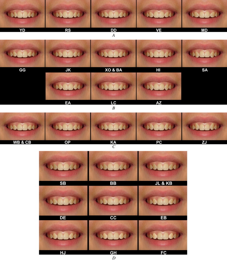
A photograph of a patient’s smile was selected that exhibited good dental alignment, having had maxillary canines substituted for missing lateral incisors. The photograph showed the maxillary teeth, lips, and surrounding skin, but the nose and chin were cropped out to reduce the confounding variables. A bilaterally symmetrical image was digitally created (Photoshop CS2 software, Adobe Systems, San Jose, Calif). The photograph was then digitally altered to create 31 images divided into 4 groups. In each group of images, 1 variable was incrementally altered; the variables were canine width (6 images), canine crown height and tip morphology (9 images), canine gingival margin height (6 images), and canine shade (10 images). A duplicate of 1 image was included in each group of images to assess intraexaminer reliability ( Fig 1 ).
The images were printed on color photographic paper in size 6 × 4 in, and each image had a unique identifier number on the reverse side of the image. The images were laid out on a table in their 4 groups in random order.
All observers in the study were given the same verbal instructions by the same examiner (E.B.). The observers were asked to study 1 group of photos at a time and then to rank them for attractiveness, from most to least attractive.
Once they had ranked the 4 groups of images and were happy with their decisions, the 4 most attractive images they selected were grouped together, and the 4 least attractive images were grouped together separately. They were then asked to pick the most attractive image from the 4 attractive images and the least attractive image from the 4 least attractive images.
Statistical analysis
The data were analyzed by using descriptive, mixed logistic regression analysis. Data analysis was performed with a statistical package (version 9, Stata, StataCorp, College Station, Tex).
Preference scores per image were computed overall and for each observer and professional group. Average ranks for each parameter were investigated along with differences in ranks given by each group.
Mixed logistic regressions were used to assess what influenced the choice for the most and the least attractive images. The independent variables were sex, age, ethnicity, amount of alteration, and professional status (group) of the observer.
Results
Each of the 4 groups of images was analyzed individually. The data were analyzed for image preference related to the person, the observer group, and the overall total.
In the overall data of canine width alterations ( Table II ), the image that was favored by most observers was image 5; 50% of the observers found it the most attractive, followed by images 1 and 6. The least attractive image was image 4, 71.67% of the observers finding it the least attractive.
| Image | Alteration | Mean score | SD | Orthodontists | Dentists | Laypeople | ||||||
|---|---|---|---|---|---|---|---|---|---|---|---|---|
| Mean | SD | Rank | Mean | SD | Rank | Mean | SD | Rank | ||||
| 1 (D.D.) | Original image | 2.46 | 1.20 | 2.08 | 0.86 | 2 | 2.33 | 0.83 | 2 | 2.98 | 1.59 | 2 |
| 2 (V.E.) | 1.5 mm wider | 4.12 | 1.20 | 4.23 | 0.95 | 4 | 4.10 | 1.24 | 4 | 4.03 | 1.39 | 5 |
| 3 (L.B.) | 1.5 mm wider (copy of image 2) | 3.98 | 1.30 | 4.35 | 0.86 | 5 | 4.20 | 0.99 | 5 | 3.38 | 1.69 | 4 |
| 4 (M.D.) | 3.0 mm wider | 5.47 | 1.00 | 5.93 | 0.35 | 6 | 5.83 | 0.45 | 6 | 4.65 | 1.29 | 6 |
| 5 (R.S.) | 1.5 mm narrower | 1.94 | 1.31 | 1.48 | 0.64 | 1 | 1.58 | 0.87 | 1 | 2.78 | 1.73 | 1 |
| 6 (Y.D.) | 3.0 mm narrower | 3.04 | 1.45 | 2.95 | 1.06 | 3 | 2.98 | 1.37 | 3 | 3.20 | 1.84 | 3 |
There were no significant differences in participant preferences (most or least attractive) related to age, sex, or ethnicity.
In the data for group-specific differences, there were significant differences in the preferences for attractiveness of the photographs comparing the orthodontists and the laypeople, and the dentists and the laypeople (there were no significant differences between the orthodontists and dentists).
By using multivariate logistic regression, both the orthodontist and the dentists were statistically significantly more likely to pick image 5 as attractive (ranked 1 or 2). Orthodontists and dentists were 12.5 and 7.7 ( P = 0.001) times more likely, respectively, to pick image 5 as attractive; 92.5% of the orthodontists, 85% of the dentists, and 58% of the laypeople ranked image 5 as 1 or 2.
The least attractive image was number 4. It was almost unanimously ranked as the least attractive image by all groups; 72% of all observers found image 4 the least attractive. Both orthodontists and dentists were more likely than laypeople to rank this image least attractive; 95% of the orthodontists and 85% of the dentists ranked it least attractive, but only 35% of the laypeople agreed with this. No observer chose it as the most attractive. It was never chosen in second place, either by the dentists or the orthodontists and only by 5% of the laypeople.
All 3 groups were in general agreement about which images were attractive and unattractive, in terms of width of the canine. However, the spread of the data and therefore the variations in opinions of the laypeople were greater than those of the orthodontists and dentists.
In the overall data of canine tip alterations ( Table III ), the image that was favored by most observers was image 1; 35.8% of them found it the most attractive, followed by images 3, 4, and 2. The least attractive image was image 9; 57.5% of the observers found it the least attractive image.
| Image | Altered canine tip | Mean score | SD | Orthodontists | Dentists | Laypeople | ||||||
|---|---|---|---|---|---|---|---|---|---|---|---|---|
| Mean | SD | Rank | Mean | SD | Rank | Mean | SD | Rank | ||||
| 1 (J.K.) | Original image | 2.76 | 1.98 | 1.88 | 1.2 | 1 | 2.69 | 2.08 | 1 | 3.68 | 2.13 | 3 |
| 2 (X.O.) | Increased 0.5 mm | 3.66 | 2.3 | 2.45 | 1.38 | 2 | 3.56 | 1.94 | 4 | 4.93 | 2.67 | 5 |
| 3 (B.A.) | Increased 0.5 mm (copy of image 2) | 3.28 | 1.84 | 3.18 | 1.28 | 3 | 3.13 | 1.88 | 2 | 3.54 | 2.25 | 1 |
| 4 (G.G.) | Reduced 0.5 mm | 3.55 | 2.03 | 3.28 | 1.34 | 4 | 3.33 | 1.9 | 3 | 4.02 | 2.6 | 4 |
| 5 (H.I.) | Increased 1.0 mm | 4.73 | 1.98 | 5.30 | 1.11 | 5 | 5.28 | 1.82 | 5 | 3.66 | 2.34 | 2 |
| 6 (E.A.) | Reduced 0.5 mm and pointed | 6.69 | 1.65 | 7.20 | 1.04 | 7 | 6.59 | 1.77 | 7 | 6.29 | 1.9 | 8 |
| 7 (S.A.) | Increased 1.5 mm | 5.72 | 1.75 | 5.48 | 1.36 | 6 | 5.82 | 1.7 | 6 | 5.85 | 2.13 | 6 |
| 8 (L.C.) | Increased 0.5 mm and pointed | 6.88 | 1.81 | 7.48 | 0.99 | 8 | 7.10 | 1.79 | 8 | 6.1 | 2.18 | 7 |
| 9 (A.Z.) | Increased 1.0 mm and pointed | 7.73 | 2.07 | 8.78 | 0.53 | 9 | 7.49 | 2.35 | 9 | 6.93 | 2.32 | 9 |
There were no significant differences in observer preferences related to age, sex, or ethnicity.
In the multivariate logistic regression, the orthodontists and dentists were more likely than laypeople to find image 1 attractive and rank it either as 1 or 2. The odds of this were 7.14 times greater for orthodontists than laypeople ( P = 0.000) and 4 times greater for dentists than laypeople ( P = 0.01).
Age ( P = 0.97), sex ( P = 0.20), and ethnicity ( P = 0.99) had no significant effect on attractiveness rank. Both orthodontists and dentists were more likely than laypeople to rank image 1 as most attractive. Orthodontists were 3.71 times more likely than laypeople ( P = 0.02) and dentists were 1.85 times more likely than laypeople ( P = 0.26) to prefer image 1.
The image found least attractive more often was image 9; 57.5% of all observers found image 9 the most unattractive. It was chosen by 82.5% of the orthodontists, 51.2% of the dentists, and 40% of the laypeople. No orthodontist or layperson observer chose it as the most attractive.
Orthodontists were significantly more likely than dentists ( P = 0.003) and laypeople ( P = 0.0000) to pick image 9 as the least attractive.
In the overall data of canine shade ( Table IV ), the image that was favored by most observers was image 3 (10 times brighter); 23.33% found it to be the most attractive, followed by image 2 (5 times brighter), image 4 (15 times brighter), and image 1 (original image). The least attractive image was image 10 (20 times darker), the darkest image; 78.33% of the observers found it the least attractive image. No participant scored image 10 as the most attractive.
| Image | Alteration | Mean score | SD | Orthodontists | Dentists | Laypeople | ||||||
|---|---|---|---|---|---|---|---|---|---|---|---|---|
| Mean | SD | Rank | Mean | SD | Rank | Mean | SD | Rank | ||||
| 1 (C.C.) | Original | 3.77 | 1.83 | 3.60 | 1.46 | 4 | 3.28 | 1.68 | 2 | 4.43 | 2.12 | 5 |
| 2 (E.B.) | 5 times brighter | 3.53 | 2.32 | 2.95 | 2.16 | 2 | 3.25 | 2.16 | 1 | 4.38 | 2.43 | 4 |
| 3 (H.J.) | 10 times brighter | 3.17 | 2.12 | 2.73 | 1.71 | 1 | 3.35 | 2.39 | 3 | 3.43 | 2.18 | 1 |
| 4 (G.H.) | 15 times brighter | 3.72 | 2.28 | 3.20 | 1.57 | 3 | 4.38 | 2.6 | 4 | 3.58 | 2.44 | 2 |
| 5 (F.C.) | 20 times brighter | 4.75 | 2.40 | 4.95 | 2.17 | 6 | 5.33 | 2.07 | 6 | 3.98 | 2.76 | 3 |
| 6 (D.E.) | 5 times darker | 5.06 | 1.88 | 4.68 | 1.75 | 7 | 4.98 | 1.95 | 5 | 5.53 | 1.88 | 6 |
| 7 (J.L.) | 10 times darker | 6.33 | 1.97 | 6.98 | 1.48 | 5 | 6.18 | 1.78 | 7 | 5.85 | 2.40 | 7 |
| 8 (K.B.) | 10 times darker (copy of image 7) | 7.06 | 1.83 | 7.38 | 0.98 | 8 | 6.68 | 1.95 | 8 | 7.13 | 2.28 | 8 |
| 9 (B.B.) | 15 times darker | 8.18 | 1.92 | 8.80 | 0.85 | 9 | 8.33 | 2.14 | 9 | 7.43 | 2.23 | 9 |
| 10 (S.B.) | 20 times darker | 9.44 | 1.44 | 9.75 | 0.74 | 10 | 9.28 | 1.92 | 10 | 9.3 | 1.38 | 10 |
Stay updated, free dental videos. Join our Telegram channel
VIDEdental - Online dental courses


