APPENDIX c CAD/CAM Computed Tomography
CD-ROM HANDLING REQUIREMENTS
The following are the CD-ROM handling requirements for all bone modeling exams sent to Techmedica.
| CT Scan Protocols | Data Transmission | Procedure |
|---|---|---|
| Patient movement | Scanner maintenance check | Remove all metal from patient’s head and neck (e.g., jewelry, hairpins). |
| Anatomic feature | Mandible | Position the patient with the head as far into the head holder as possible. Wear gloves. |
| Patient positioning | With the patient lying supine, place the head as far as possible into the coronal head holder so that the mandible is behind the metal in the forward part of the head holder. Place a pillow under the patient’s knees for comfort. Wedge sponges between the head and head holder, if space is available, to prevent patient movement. Strap the chin and forehead. | Wet the intraoral jig and shake off excess moisture. Sprinkle the gum areas of the jig with Rigident powder and shake off excess. Insert the jig into the patient’s mouth. Lubrication of the lips with lubricating jelly may be necessary. Attach the vacuum line to the jib tube and set on the lowest possible setting that still evacuates saliva. If the vacuum “grabs” the tongue, reduce vacuum. Palpate the inferior border of the anterior body of the mandible and position it parallel to the scan plane. Secure patient’s head in head holder. |
| Plastic rod location | Tape the ⅜ -inch diameter plastic rod to the patient’s face, down the midline, from the end of the nose to past the end of the chin, so that the rod is approximately perpendicular to the scan plane. |
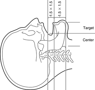 |
(GE 8800 scanner)
(GE 9800 scanner)
Stay updated, free dental videos. Join our Telegram channel
VIDEdental - Online dental courses


