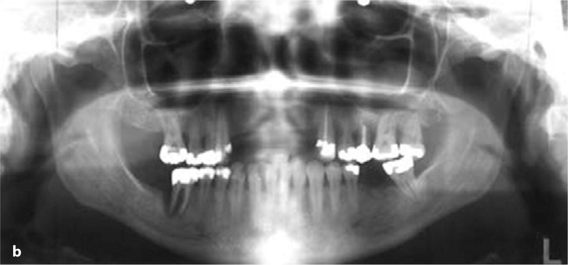
Fig. 14.1a,b.
Teleradiograph and OPG of the preoperative situation. All teeth in the maxilla except for tooth 13 were extracted. All maxillary molars exhibit tunnelling defects. Extraction of tooth 46 is indicated as well. Surgical pre-treatment by an orthodontist was not considered useful. The ANB was about –4 degrees. The skeletal consequences of prognathism do not per se cause any problems
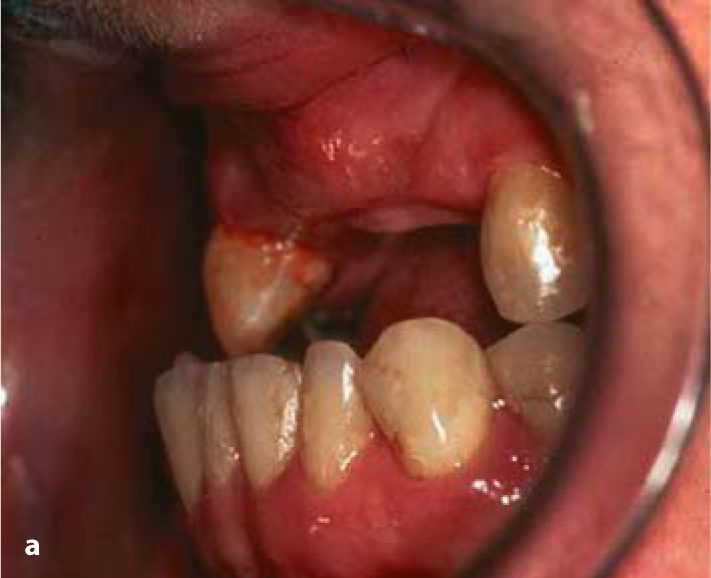
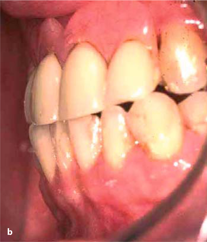
Fig. 14.2a,b.
The anterior teeth in the maxilla were extracted and replaced by a “corrective” ceramo-metal denture for aesthetic reasons. This denture was actually the source of the periodontal problems diagnosed before implant treatment
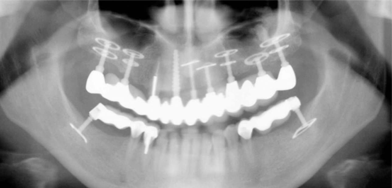
Fig. 14.3.
A total of seven BOI implants and one compression screw were inserted. Tooth 13 was also included in the restoration. This OPG was taken 5 years after the maxilla had been restored. The bone structures around the load-transmitting surfaces do not reveal any deficits in terms of healing. No crestal bone is apparent. The treatment concept in this patient was based on multiple rather than strategic implant placement. In the meantime, the mandible was restored as well
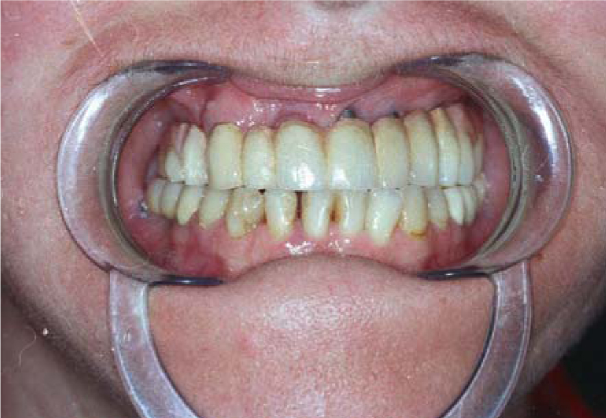
Fig. 14.4.
Clinical situation after 6 years
Even if the maxilla is strongly retruded, an attempt should be made to establish correct relations between the canines and to insert the maxillary anterior implants at the canine position.
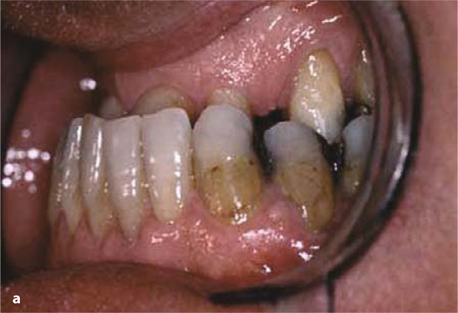
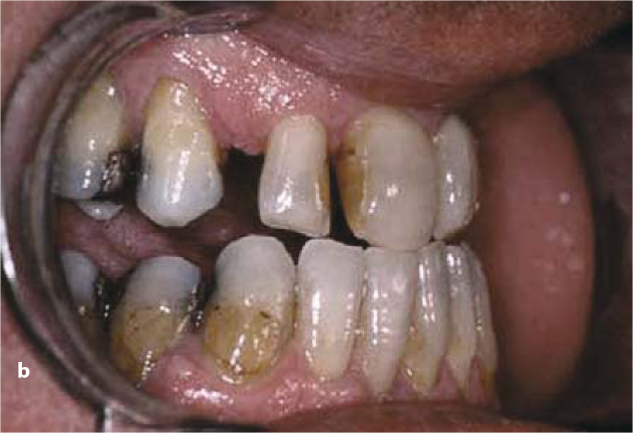
Fig. 14.5a,b.
Intraoral view of the residual dentition in the maxilla. Tooth 22 was congenitally missing. While the patient was able to establish an end-to-end occlusion, habitual chewing was only possible in a crossbite configuration
Stay updated, free dental videos. Join our Telegram channel

VIDEdental - Online dental courses


