Fig. 23.1
Traces of tooth marks on crown caps of beer bottles (Solheim 1980)

Fig. 23.2
Trace comparison (comparative microscopy: crown cap of a beer bottle (left – right)). Many indentations show conformity
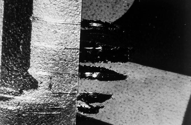
Fig. 23.3
Trace comparison (at higher magnification): the microfurrow on the crown cap of the mineral water bottle (left) and the beer bottle (right) match exactly
However, teeth may also leave marks during a fall or because of a punch against the teeth. In such cases, the teeth often are damaged or fractured. Then, the scene must be searched for the dropped or fractured teeth. Most often, tooth marks occur in the skin (Ligthelm and van Niekerk 1995). The skin can be cut and/or a piece of skin could be bitten out (Graw et al. 1998). In a conflict, both parties, perpetrator and victim, may have tooth marks on the skin.
Particularly in the investigation of sexual crimes, bodily injuries, and other violent crimes, bite marks can be found on the body of the injured person and, as a result of defensive actions, also on the body of the perpetrator (Prokop 1966; Free 1995; Graw et al. 1998). In all cases where bite marks are found, attention must also be paid to traces of blood and/or saliva. This is useful for the reconstruction of the course of the crime and to determine any possible intoxication of the perpetrator (Zerndt and Simon 1962).
In cases of rape, tooth marks (only with impressions and subepithelial hemorrhage) often resemble a love bite. If the victim survives the attack, these marks disappear after few hours. Therefore, the tooth marks must be examined as quickly as possible. Subepithelial bleeding is still visible after several days and may become even more obvious. Therefore, the victim should be examined on several consecutive days. In addition, an investigation under infrared or ultraviolet light makes the marks more obvious (Ruddick 1974). However, subepithelial bleeding only shows little detail and is of limited importance. Frequently found are bites with ring-shaped subepithelial bleeding as a result of a suction process. The dimensions of the bite may change (Thompson and Phillips 1994).
Unlike animals, humans suck more often than they bite. Animals do not leave central subepithelial hematomas between the dental arches. In case of a suspected animal bite, the investigation and bite comparison should be done in cooperation with a veterinarian (Ligthelm and Niekerk 1995).
The mechanics of bite injuries with cuts of the epidermis, “pull bite” (Fig. 23.4), is fundamentally different from the “suck bite” (Fig. 23.5), with enrichment of the epidermis in front of the cutting edges of the teeth (Pilz et al. 1980). Experimenting with suction cups on the skin (Fig. 23.6) produces a circular mark that develops into an oval shape after removal of the suction cup (Pilz et al. 1980). This behavior corresponds to the cleavage lines, as described by Langer (1861).
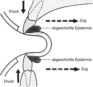
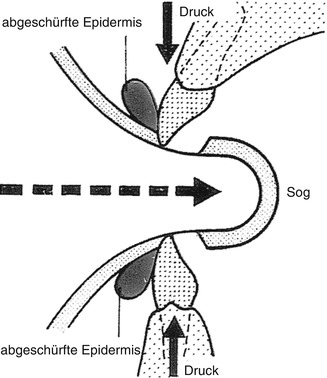
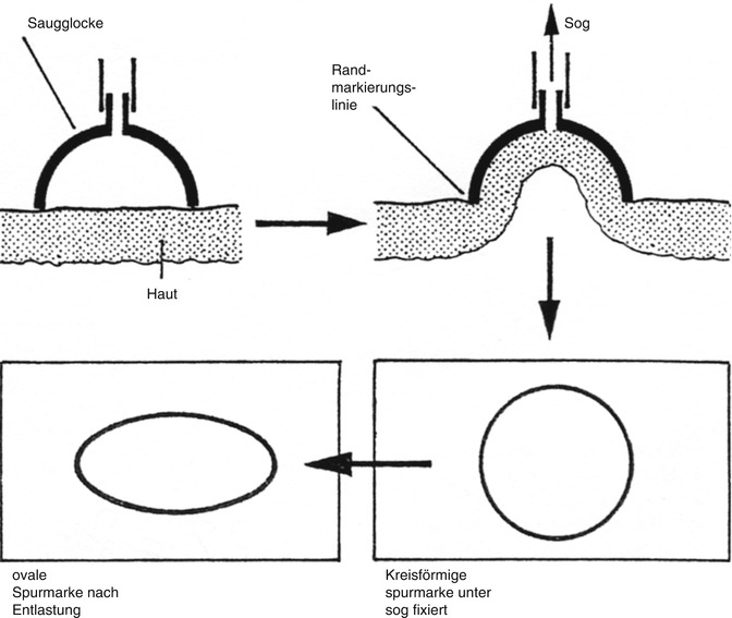
Fig. 23.4
Mechanics of the bite injury with cut of the epidermis by the “distortion-pull bite”
Fig. 23.5
Mechanics of “suck bite” with “enrichment” of the epidermis (Modified after Zerndt, with courtesy of Prof. Dr. W. Pilz, Arnstadt)
Fig. 23.6
Experimental suction marks on the skin (Courtesy of Prof. Dr. W. Pilz, Arnstadt)
The nature of the force acting on the skin, i.e., whether the bite was made with “burrowing” movements or if the skin was in a stretched or warped condition, is also important. In this case, moving into a relaxed state also shifts the bite mark. This is particularly true for arms and legs (surveys on human volunteers), and is another uncontrollable unknown in the analysis of bite marks (Pilz et al. 1980).
In order to test the validity of evaluations of bite marks, Pilz et al. (1980) conducted bite mark comparisons (native trace setting in the human skin) on different models in a blind test. The accuracy of the evaluation by photographs was very dependent on the time when the photos were taken. The evaluation of the 24-h model showed a precision value of only 9 %! A high percentage of accuracy is achieved when the bite mark provides clear imprints and is photographed as soon as possible.
Nehrkorn and Sturm (1974) studied the problem of whether:
1.
Typelike normal teeth have recognizably different impressions at all
2.
Active bites into vital, responsive, and movable substrate are comparable to bite mark copies.
In typical normal teeth, comparisons are worthless because pseudo-identities bear the risk of false positive statements. In case of congruence only, the reference “not to be excluded” is allowed, with reference to the high rate of nonspecific similarity in typical teeth.
Abused children often have tooth marks in the skin (Fulton 1984). These traces only show impressions and subepithelial bleeding, especially if the perpetrator had no intention to kill. If the examination of the child takes place after several days, the marks usually are no longer visible and thus are hardly suitable as evidence.
Bite marks on the same object can be single or multiple, and may be spread over various regions of the body, so the entire body must be examined. Furthermore, the traces can be caused by one or several perpetrators (Pilz et al. 1980).
The localization of bites (frequency) shows that the fingers are most commonly involved (due to defensive reactions), followed by the female mammary gland (sexual crimes), and the extremities. The neck and torso are relatively rarely involved (Ulrich 1963). The locations of bites with respect to their frequencies, from most to least frequent, are (Ulrich 1963): fingers, breasts, legs, nose, hands, ears, cheeks, lips, neck, chin, shoulders, abdomen, genitals, and armpit.
A further study on the frequency of bite marks in homicides regarding the sex of the perpetrator (trace causer) and the sex of the victim (trace carrier) clearly shows that the male sex is in the majority, while the female gender predominates as a trace carrier (victim) (Ulrich 1963).
23.3 Examination Methods
Pathologically altered or dentally treated dentures are more useful for an assessment of bite marks than “normal teeth.” Note that the mechanics of the bite must be considered (Pilz et al. 1980; Sweet 1995; Zerndt 1964):
-
Simple hack bite
-
Tear or pull bite
-
Raising of soft tissues that are grasped with the teeth
-
Intensity and location of the bite
-
The vital reactions
-
The nature of the affected tissue
-
The respective base
Nevertheless, many details must be minded to avoid faulty assessments. In this regard, it is advantageous that “normal teeth” appear less than pathologically altered dentures.
The examination of bite marks in violent crimes requires great expertise. The dental expert must be included in the investigation as early as possible. The dental assessment must be stated in an opinion that is justified by proper scientific methods. Even the securing of evidence bears the risk of incorrect assessments. All traces at the scene must be secured, even the smallest details can be of great importance for the identification of the perpetrator. Under the principle of “do not touch anything, do not change anything,” the crime scene must be protected against influences that may alter the traces. False or fictitious traces can be caused by the course of the crime or may be targeted by the perpetrator. Such traces must be immediately secured (Hennis et al. 1981a).
The examination of bite/tooth marks requires interdisciplinary teamwork. Detectives, forensic scientists, serologists, and dentists are involved in the detection, securing, and evaluation of traces. An experienced dentist should be consulted in all cases where bite/tooth marks are present (Endris 1985; Sweet 1995).
Searching the literature on legal medicine, it becomes obvious that bite injuries have had greater importance in court only since a relatively short time. Meanwhile, numerous authors have studied the possibilities of identifying bites as well as the resulting conclusions about the identity of the perpetrator (Selle 1966). For the legal assessment, it is very important that bites are examined as early as possible, preferably by an experienced forensic odontologist.
Graw et al. (1998) reported a morphological finding survey on a piece of skin and its DNA typing, which led to the identification of the perpetrator. A 35-year-old woman who suffered from psychosis was found dead with multiple stab wounds. A piece of horny skin (~1 cm) was found in her oral cavity (sublingual). It could not be assigned to one of the numerous skin defects. Several men of her connections were potentially suspected and had to undergo a physical examination. One of them had a healing injury at the bending side of his left ring finger. The piece of horny skin had the contours and size and papillary lines of this wound. The DNA testing showed that the piece of skin could only be assigned to the suspect, who thereupon made a confession. The specific molecular biological study of the washed piece of skin showed a statistically computable frequency of 1:700,000 (matching feature pattern).
The following case studies are from a homicide in which the perpetrator claimed not to have perceived any bites. During the autopsy, a picture of the side view of the forehead was taken (Fig. 23.7, see arrow) and showed a bow-shaped, scratch-like cut of the epidermis between the eyebrows (the other injuries of the skin, caused by a knife, were irrelevant). Initially it was assumed that the wound was caused by a broken milk bottle. The forensic odontologist supposed that the bow-shaped forehead injury was caused by a bite of the perpetrator.
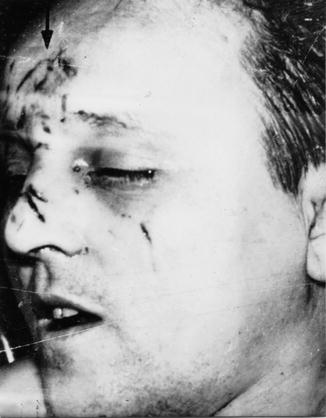
Fig. 23.7
Photography of the wound
The comparison with the plaster of the upper jaw of the perpetrator showed that the injury was caused by the right canine, all incisors, the left canine, and the anterior premolar. The width of the wound and also the width of the incisor was 7 mm. Remarkable is that the impression of the first right incisor is slightly inward (Figs. 23.8 and 23.9, arrow A). Those studies confirmed that the bow-shaped skin lesion was caused by a bite of the suspect (Rötzscher 1972). The tooth mark is more likely an impression than an active bite that emerged through the impact of the teeth of the perpetrator on the forehead of his victim, without the intent to bite. This could be explained by a violent impact (head butt) of the upper dental arch (with open mouth) during the fight with the defending victim.
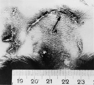
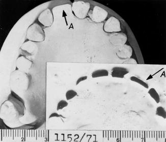
Fig. 23.8
Image of the forehead with a bow-shaped injury
Fig. 23.9
Upper jaw plaster model of the suspect with the cutout plasticine impression
An important reference criterion is an acceptable thickness of the soft tissue in the forehead area. In the area of the scalp and the soft tissues of the forehead, there is little deviation of the thickness, as the scalp, on average, is about 5- to 7-mm thick. The measurement of the soft tissue thickness according to Helmer (1998) is determined by sex, age, and type of constitution:
1.
Measuring point 17: stm — middle of the forehead, point in the center of the upper orbital margin on the line of frontotemporal to metopion
2.
Measuring point 30: mt — M. temporalis above zygion, at the height of the ectoconchion
3.
Measuring point 31: zy — zygion (the most protruding point on the cheek bone)
In the presence of an anomaly, its characteristics are valuable in identifying the bite/tooth mark. The morphological assessment has priority over the measurements of distance and angle, which only have complementary functions (Hennis et al. 1981b, c).
Prerequisite for a judicial assessment of bites is accurate anatomical knowledge of the shape and appearance of the wounds as well as profound knowledge of the dentition and the bite behavior of animals in order to determine whether the bite injury was caused by an animal or by a human (Endris 1979).
23.4 Traces Caused by Animals
Animals bite (at least during the attack) indiscriminately into covered and uncovered parts of the body. Humans usually bite into bare or bared body parts: fingers, breasts, arms, legs, nose, hands, ears, cheeks, lips, neck, jaw, shoulders, abdomen, genitals, and armpits. In particular, the fingers, ears or the nose may completely or partially be bitten off. Animals that may become a danger because of their bites are primarily pets such as dogs, cats, and horses. The owner is liable for the legal consequences of animal bites. Because many bite injuries by animals are serious and even lethal, these cases are subject to criminal law. In the USA, an estimated two to three million dog bites occur each year (60 % of the victims are children). Approximately 20 children in the USA die each year from dog bites (Berk et al. 1997).
Injuries caused by bites of nondomestic animals, such as rats, mice, wild cats, and other predators, are important during an investigation because the time of the bite (intravital or postmortem) may help to solve a crime. Postmortem injuries caused by wild animals, even smaller animals, may be confusing because they massively alter the condition of the body and must be carefully examined in order to detect whether the injuries were caused intravital or postmortem.
Tsokos and Schulz (1999) reported two cases with postmortal injuries by domestic animals that mangled their owners who had died natural deaths. The owner of two pitbull terriers laid dead and dressed in his house. The two dogs mangled his head and neck region; the larynx, the thyroid gland, parts of the carotid artery of both sides and the upper part of the esophagus and the trachea could not be found at the scene.
A 41-year-old woman had died from an acute pneumonia and laid dressed in her house. Rats gnawed her neck and upper thorax. The larynx, the thyroid gland, parts of the carotid artery of both sides and the upper part of the esophagus and the trachea could not be found at the scene. Rat bites are also found on living persons. This etiological difficulty increases the significance of the cases.
Strauch (1927) studied cases of bite injuries by cats: “Felids are markedly predators which put the main emphasis on the hunt of living, warm prey.” Nevertheless, there are cases where cats ate dead bodies. Commencing or advanced decay does not keep them from the corpse.
In identification of animal bites it is difficult to recognize a bite because most of the tissue is destroyed by tearing bites and, thus, it is difficult to detect individual features. Houtrow (1963) reported a case of a 14-year-old boy who was found dead in a park in Vienna, maimed by injuries that seemed to be stab wounds. Investigations and the forensic autopsy revealed that the injuries were from several German Shepherd dogs who had largely mangled the body.
Comparative studies of skulls of dogs (German shepherd, Boxer) and a polar bear (Du Chesne and Rötzscher 1968) provided both surprising similarities and differences between the species and the sets of their bites. The dentition of the Boxer shows physiological prognathism (reverse front bite) with expected bite ratios strains and bruises in the tissue. In the German shepherd, the front teeth rows slide like scissor branches close to each other. In dog bites, the relatively long canines (carnassials) leave impressions that are similar to injuries (Figs. 23.10 and 23.11) by conical objects (Pilz et al. 1980).
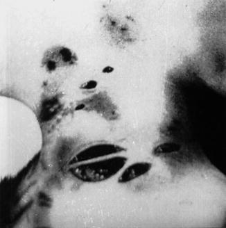
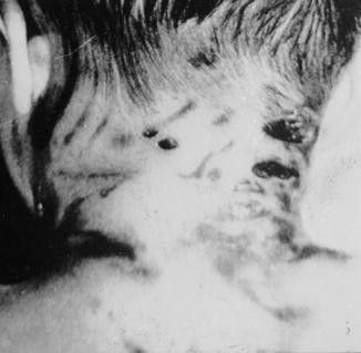
Fig. 23.10
Animal bite (shepherd dog), similar to stab wounds
Fig. 23.11
Bite wounds; tooth marks, slide traces, and subdermal bleedings
Conclusions about the nature and course of the injury can only be drawn by comparative analysis of teeth and bite injuries. For example, the Forensic Institute in Leipzig had to examine the body of a 2.5-year-old boy who had fallen over the railing into the pool of a polar bear compound. First the child was submerged and brought up into the paws of the polar bear. He brought the child back to land where he was rescued by the guards after 10–20 min. The child died on the way to the hospital. The autopsy showed numerous bites, abrasion injuries, and scratches on head and body. The pericranium was scalped. The right lower leg had a characteristic ring-shaped discoloration of the skin with abrasion injuries. It was assumed that the polar bear pressed his slightly opened mouth against the skin and sucked. This type of bite was interpreted for the playful behavior of the polar bear during the accident. It would have been easy for the polar bear to mangle the child’s leg (Du Chesne 1968). Note that the main danger of animal bites is less the injury itself as its infectivity.
Minor injuries are underestimated by the patient and can be starting points of infections, such as pasteurellosis (Pasteurella multocida and septica), septicopyemia (Streptobacillus moniliformis), Sodoku (“rat bite fever”), and Weil’s disease (Leptospira icterohaemorrhagiae) (Mollaret 1969).
Tsokos and Schulz (1999) compiled literature about bites by domestic animals and their localization; this information is presented in Chart 23.1.
Chart 23.1
Domestic animal bites and their locations in a literature collection
|
Authors
|
Age/gender
|
Cause of death
|
Animal
|
Time between death and postmortem injuries
|
Location
|
Traces of blood
|
|---|---|---|---|---|---|---|
|
Böhmer (1925)
|
Newborn/(u)
|
(u)
|
Mouse
|
(u)
|
Extremities
|
(u)
|
|
Böhmer (1925)
|
3 days/m
|
Cerebral hemorrhage
|
Rats
|
3 days
|
Face
|
No
|
|
Mttmeyer et al. (1976)
|
49 years/m
|
Cerebral hemorrhage
|
Dog
|
(u)
|
Penis
|
On carpet
|
|
Mätzler (1977)
|
66 years/m
|
Natural death
|
Dog (poodle)
|
(u)
|
Abdomen, penis, anus
|
No
|
|
Weiler (1978)
|
77 years/m
|
Pneumonia
|
Dog
|
8 days
|
Abdomen, pelvis, penis
|
No
|
|
Pötsch-Schneider and Endris (1984)
|
53 years/m
|
Cerebral hemorrhage
|
Dog
|
(u)
|
Decapitation
|
Large amounts on head and neck
|
|
Haglund (1992)
|
27 years/m
|
Suicide by propane gas inhalation
|
Rats
|
3 days
|
Skeletonization of the head and (u)
|
(u)
|
|
Hayase et al. (1994)
|
59 years/m
|
Ischemic heart disease
|
Dog
|
3 days
|
Front chest wall was missing, internal organs intact
|
no
|
|
Rossi et al. (1994)
|
53 years/m
|
Pneumonia, diabetes, heart disease
|
Dog (German shepherd)
|
(u)
|
Skeletonization of head and neck, internal organs missing
|
Tracks
|
|
Rossi et al. (1994)
|
82 years/m
|
Myocardial ischemia
|
Dog (German shepherd)
|
(u)
|
Skeletonization of head and neck, internal organs partially missing
|
Tracks
|
|
Rossi et al. (1994)
|
42 years/f
|
Pulmonary edema, alcohol intoxication
|
Dog (Setter)
|
16 h
|
Face
|
(u)
|
|
Rossi et al. (1994)
|
32 years/m
|
Suicide by intoxication
|
Stay updated, free dental videos. Join our Telegram channel
VIDEdental - Online dental courses


