Introduction
The purposes of this retrospective study were to investigate the apical root resorption of maxillary incisors in orthodontic patients with en-masse maxillary anterior retraction and intrusion with miniscrews and the factors disposing a patient to apical root resorption.
Methods
Fifty adult patients with maxillary protrusion were included; 30 were treated with miniscrews and extraction of the maxillary first premolars (group I), and 20 were treated with extraction of the maxillary first premolars (group II). For each patient, periapical films of the maxillary incisors and lateral cephalometric radiographs were taken before and after treatment to evaluate apical root resorption and cephalometric measurements. The intergroup differences were analyzed with the Student t test and the correlations between apical root resorption and cephalometric measurements were analyzed by the Pearson correlation.
Results
The apical root resorption values were 16.0% to 20.0% (2.5-2.8 mm) in group I and 13.4% to 14.4% (2.1-2.3 mm) of the original root length in group II. Group I had significantly more severe Class II jaw discrepancy (ANB, 7.1° ± 1.9°) than did group II (ANB, 3.2° ± 2.9°). The amount of maxillary en-masse anterior retraction (8.2 ± 2.4 mm), the duration of treatment (28.3 ± 7.3 months), and apical root resorption of maxillary lateral incisors were significantly greater in group I than in group II. Apical root resorption of the maxillary central incisors was significantly correlated to the duration of treatment but not to the amount of en-masse retraction, intrusion, or palatal tipping of maxillary incisors.
Conclusions
Miniscrew anchorage allows for more maxillary en-masse anterior retraction in patients with severe Class II cases. But the time needed for the greater amount of maxillary en-masse anterior retraction with miniscrew anchorage is longer and might dispose the patient to more apical root resorption.
The growing demand for orthodontic treatment methods that require minimal compliance and maximal anchorage control, particularly by adults, has led to the expansion of implant technology. Miniscrews have been introduced as temporary anchorage devices for various purposes: canine retraction, anterior retraction, en-masse anterior retraction, molar uprighting, distalization, and protraction. They have the advantages of smaller size, more implant sites and indications, simpler placement surgically and connection orthodontically, short or even no waiting period, no need for laboratory work, easier removal after treatment, and lower cost than implants, onplants, and miniplates.
Without a limit in anchorage, we can now move teeth farther, position the maxillary or mandibular incisors in ideal positions and inclinations, and intrude the incisors farther with miniscrews. Nevertheless, the roots of maxillary or mandibular incisors would travel farther through the dentoalveolus, and treatment could also be longer than with conventional approaches. All of these are considered predisposing factors for apical root resorption. However, apical root resorption in patients with miniscrew anchorage has not been studied thoroughly yet.
The purposes of this retrospective study were to study the apical root resorption of maxillary incisors in orthodontic patients with en-masse maxillary anterior retraction and intrusion with miniscrews and the factors disposing them to apical root resorption.
Material and methods
Fifty adults who had their maxillary first premolars extracted and en-masse maxillary anterior retraction and intrusion of their maxillary dentoalveolar protrusion, and had clear and readable pretreatment (T1) and posttreatment (T2) periapical and lateral cephalometric radiographs were selected from our database ( Table I ). They had no medical history of asthma, hypothyroidism, or maxillofacial trauma, and no dental history of root canal treatment of the maxillary incisors or previous orthodontic treatment. Thirty patients were treated with miniscrew anchorage and extraction of the maxillary first premolars (group I), and 20 were treated with extraction of the maxillary first premolars without miniscrew anchorage (group II). The group II patients were treated before orthodontic miniscrews were available.
| n | Age (y) | |||||
|---|---|---|---|---|---|---|
| Group | Men | Women | Mean | SD | Minimum | Maximum |
| I | 0 | 30 | 26.5 | 5.5 | 20.2 | 44.6 |
| II | 4 | 16 | 22.5 | 1.6 | 20.3 | 26.1 |
| Total | 4 | 46 | 25.4 | 5.6 | 20.2 | 44.6 |
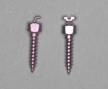
The miniscrews were the Lin/Liou orthodontic mini anchor system (Mondeal Medical System GMBH, Tuttlingen, Germany). The size was 2 mm in diameter and 9 mm in length ( Fig 1 ). The miniscrews were placed in the infrazygomatic crest of the maxilla.

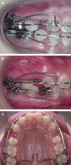
The appliances and mechanisms for the en-masse maxillary anterior retraction and intrusion are described in Figure 2 . They were the same for all patients and performed by the same orthodontist (P.M.H.C.). The forces were 250 g for en-masse anterior retraction and 100 g for intrusion.

The T1 and T2 cephalometric radiographs were traced and superimposed on the anterior cranial base and best fitted on the cranial vault. A horizontal reference line (x-axis) through sella was constructed anteriorly 7° to the sella-nasion line, and a perpendicular line through sella to the horizontal reference line was constructed as the vertical reference line (y-axis). The root shortening of the maxillary incisor at T2 from apical root resorption returned to its root length at T1 by superimposing its crown contour at T2 with the crown contour at T1.
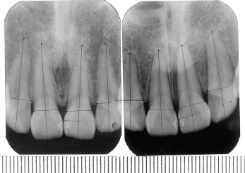
The T1 and T2 cephalometric tracings and periapical films with a ruler ( Fig 3 ) were scanned into a computer, calibrated, and measured in a 1:1 ratio with ImageJ software (version 1.37, National Institutes of Health, Bethesda, Md, http://rsb.info.nih.gov/ij/ ) for (1) maxillary incisor proclination (1-SN), SNA, SNB, and ANB angles at T1; (2) amount of anterior retraction (horizontal distance to the y-axis) and intrusion (vertical distance to the x-axis) of the incisor tip and root apex from T1 to T2; (3) palatal tipping of the maxillary incisors (1-SN-T1-2) from T1 to T2; and (4) on the periapical films, apical root resorption of the maxillary central and lateral incisors from T1 to T2.

Apical root resorption of the maxillary incisors from T1 to T2 was measured as length and percentage of shortening of the original root length at T1 by the following : apical root resorption (mm) = C1/C2 (R1 – R2); apical root resorption (%) = C1/C2 (R1 – R2)/R1 (C1, T1 radiographic incisor crown length; C2, T2 radiographic crown length; R1, T1 radiographic root length; R2, T2 radiographic root length).
Statistical analysis
Ten periapical and cephalometric radiographs were randomly selected for the study of method errors. The maxillary right central incisor crown lengths on the periapical films and the lengths of SN on the cephalometric radiographs were measured again 2 months later and analyzed with paired t tests ( P <0.05) for systemic errors, and analyzed with the formula √(ΣD 2 /2 N) for measurement errors, where D is the difference between repeated measurements and N is the number of repeated measurements.
The intergroup differences of the cephalometric measurements, duration of treatment, and apical root resorption were analyzed with the Student t test ( P <0.05). Their correlations (groups I and II combined) were analyzed with the Pearson correlation ( P <0.05).
Results
The systemic error of cephalometric measurement was 0.21 mm ( P = 0.092), and its measurement error was 0.10 mm. The systematic error of apical root resorption measurement was−0.27 mm ( P = 0.079), and its measurement error was 0.10 mm.
The pretreatment 1-SN and SNA values were similar between the groups. SNB at T1 was significantly less, and ANB was significantly greater in group I than in group II ( Table II ).
The amounts of en-masse anterior retraction of the maxillary incisor tip and root apex in group I were significantly greater than in group II. The duration of treatment of group I was significantly longer than in group II. However, the amount of intrusion of the maxillary incisor tip or root apex, and 1-SN-T1-2 were not significantly different between the groups ( Table III ).
| Group I | Group II | P value | |
|---|---|---|---|
| Retraction at incisor tip (mm) | 8.2 ± 2.4 | 6.5 ± 2.1 | 0.013 ∗ |
| Intrusion at incisor tip (mm) | 0.4 ± 2.0 | 0.0 ± 1.6 | 0.459 |
| Retraction at root apex (mm) | 3.0 ± 2.7 | 1.3 ± 1.6 | 0.018 ∗ |
| Intrusion at root apex (mm) | 2.7 ± 1.8 | 2.5 ± 1.4 | 0.709 |
| 1-SN-T1-2 (°) | 14.4 ± 10.0 | 13 .9 ± 7.5 | 0.856 |
| Duration of treatment (mo) | 28.3 ± 7.3 | 22.7 ± 5.0 | 0.003 † |
Apical root resorption of the maxillary central incisors was not significantly different between the groups, but apical root resorption of the maxillary lateral incisors was significantly greater in group I than in group II ( Table IV ).
| Maxillary incisor | Group I | Group II | P value | |
|---|---|---|---|---|
| Right lateral | (%) | 20.0 ± 7.3 | 14.4 ± 7.3 | 0.017 ∗ |
| (mm) | 2.7 ± 1.0 | 2.1 ± 1.4 | ||
| Left lateral | (%) | 19.6 ± 6.6 | 14.4 ± 8.5 | 0.030 ∗ |
| (mm) | 2.8 ± 1.0 | 2.3 ± 1.7 | ||
| Right central | (%) | 16.8 ± 8.8 | 13.6 ± 7.6 | 0.197 |
| (mm) | 2.5 ± 1.4 | 2.1 ± 1.5 | ||
| Left central | (%) | 16.0 ± 9.2 | 13.4 ± 7.3 | 0.299 |
| (mm) | 2.5 ± 1.5 | 2.1 ± 1.3 |
Stay updated, free dental videos. Join our Telegram channel

VIDEdental - Online dental courses


