Head and Neck Anatomy
Learning Outcomes
On completion of this chapter, the student will be able to achieve the following objectives:
• Pronounce, define, and spell the Key Terms.
• Identify the regions of the head.
• Locate and identify the bones of the cranium and face.
• Locate and identify the muscles of the head and neck.
• Identify and trace the routes of the blood vessels of the head and neck.
• Identify the components of the temporomandibular joint.
• Describe the action of the temporomandibular joint.
• Identify the locations of major and minor salivary glands and associated ducts.
• Describe and locate the divisions of the trigeminal nerve.
• Identify the locations of major lymph node sites of the body.
• Identify and locate the paranasal sinuses of the skull.
• Integrate knowledge about head and neck anatomy into clinical practice.
Electronic Resources
 Additional information related to content in Chapter 9 can be found on the companion Evolve Web site.
• Labeling Exercise: Identify the Bones and Landmarks of the Frontal View of the Skull
• Labeling Exercise: Identify the Bones and Landmarks of the Hard Palate
• Labeling Exercise: Identify the Major Arteries of the Face
• Labeling Exercise: Identify the Major Veins of the Face
• WebLinks
Key Terms
Alveolar process Portion of the maxillary bones that form the support for teeth of the maxillary arch.
Articular (ahr-TIK-yoo-luer) disc Cushion of dense, specialized connective tissue that divides the articular space into upper and lower compartments; also known as the meniscus.
Articular eminence Raised portion of the temporal bone just anterior to the glenoid fossa.
Articular space Space between the capsular ligament and between the surfaces of the glenoid fossa and the condyle.
Buccal (BUK-ul) Region of the head that refers to structures closest to the inner cheek.
Circumvallate lingual papillae (sir-kum-VAL-ayt LING-gwul puh-PIL-ee) Large tissue projections on the tongue.
Condyloid process The posterior process of each ramus; articulates with a fossa in the temporal bones to form the temporomandibular joint; also known as the mandibular condyle.
Coronal suture Line of articulation between the frontal bone and parietal bones.
Cranium (KRAY-nee-um) Eight bones that cover and protect the brain.
External auditory meatus Bony passage of the outer ear.
Foramen magnum Large opening in the occipital bone that connects the vertical canal and the cranial cavity.
Fossa (FOS-ah, FAW-suh) Wide, shallow depression on the lingual surfaces of anterior teeth.
Frontal Region of the head pertaining to the forehead.
Frontal process Process of the zygomatic bone that extends upward to articulate with the frontal bone at the outer edge of the orbit.
Glenoid fossa Area of the temporal bone where condyles of the mandible articulate with the skull.
Greater palatine (PA-luh-tine) nerve Nerve that serves the posterior hard palate and the posterior lingual gingiva.
Hamulus A hook-shaped process.
Infraorbital (in-fruh-OR-bi-tul) Region of the head below the orbital region.
Lacrimal (LAK-ri-mul) bones Paired facial bones that help form the medial wall of the orbit.
Lambdoid suture Line of junction between the occipital and parietal bones.
Lateral pterygoid plate Point of origin for internal and external pterygoid muscles.
Lymphadenopathy (lim-fad-uh-NOP-uh-thee) Disease or swelling of the lymph nodes.
Masseter (muh-SEE-tur) The strongest and most obvious muscle of mastication.
Mastoid process Projection on the temporal bone located behind the ear.
Maxillary tuberosity Large, rounded area on the outer surface of the maxillary bones in the area of the posterior teeth.
Meatus The external opening of a canal.
Medial pterygoid plate Plate that ends in the hook-shaped hamulus.
Mental Region of the head pertaining to or located near the chin.
Mental protuberance Part of the mandible that forms the chin.
Nasal Region of the head that pertains to or is located near the nose.
Nasal conchae Projecting structures found in each lateral wall of the nasal cavity and extending inward from the maxilla; singular, concha.
Occipital (ok-SIP-i-tul) Region of the head overlying the occipital bone and covered by the scalp.
Oral Region of the head pertaining to or located near the mouth.
Orbital Region of the head pertaining to or located around the eye.
Ossicles The bones of the middle ear.
Parietal (puh-RYE-e-tul) Pertaining to the walls of a body cavity.
Parotid (puh-ROT-id) duct Duct associated with the parotid salivary gland, which opens into the oral cavity at the parotid papilla.
Process A prominence or projection on a bone.
Pterygoid process Process of the sphenoid bone, consisting of two plates.
Sagittal suture Suture that is located at the midline of the skull, where the two parietal bones are joined.
Sphenoid sinuses Sinuses that are located in the sphenoid bone.
Sternocleidomastoid (stur-noe-klye-doe-MAS-toid) Major cervical muscle.
Styloid process Process that extends from the undersurface of the temporal bone.
Symphysis menti (SIM-fi-sis MEN-tee) The separation of the mandible at the chin that occurs at birth.
Temporal Region of the head superior to the zygomatic arch.
Temporal process Process that articulates with the zygomatic process of the temporal bone to form the zygomatic arch, which creates the prominence of the cheek.
Temporomandibular (tem-puh-roe-man-DIB-yoo-lur) joint (TMJ) Joint on each side of head that allows movement of the mandible.
Trapezius (truh-PEE-zee-us) Major cervical muscle.
Trigeminal nerve The nerve that is the primary source of innervation for the oral cavity.
Zygomatic Region of the head pertaining to or located near the zygomatic bone (cheekbone).
Zygomatic arch The arch formed when the temporal process of the zygomatic bone articulates with the zygomatic process of the temporal bone.
Zygomatic process The process of the maxillary bones that extends upward to articulate with the zygomatic bone.
In this chapter, you will learn the anatomic basis for the clinical practice of dental assisting. You will learn the names and locations of bones of the skull and face, facial nerves, lymph nodes, and salivary glands. You will identify muscles of the head and neck, including the facial muscles, which create facial expressions and help to open and close the mouth and swallow food.
You will find that knowledge of anatomic landmarks is a necessity as you begin to mount radiographs.
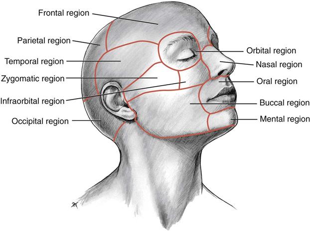
Regions of the Head
The head can be divided into 11 regions: frontal, parietal, occipital, temporal, orbital, nasal, infraorbital, zygomatic, buccal, oral, and mental. As you continue this chapter, you will encounter references to these regions of the head (Fig. 9-1).

Bones of the Skull
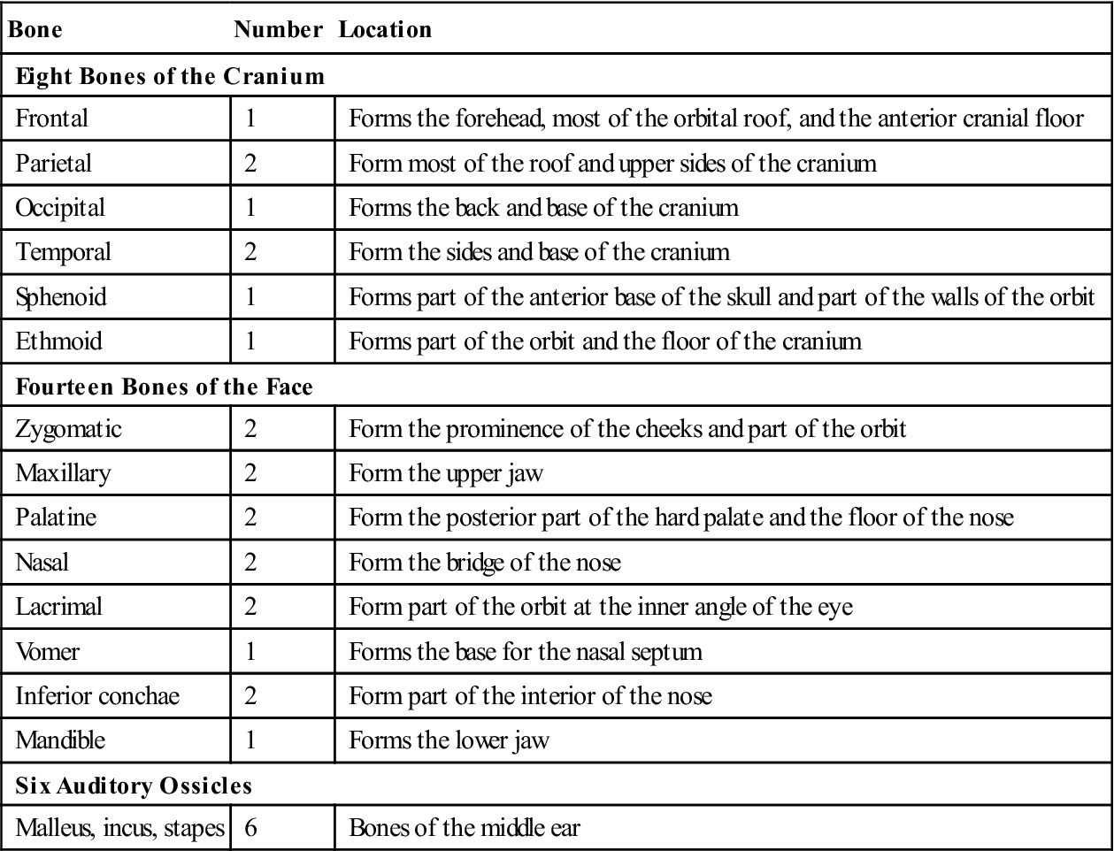
The human skull is divided into two sections: the cranium and the face. The cranium is composed of eight bones that cover and protect the brain; the face consists of 14 bones (Table 9-1). Specific anatomic terms are used to describe landmarks on these bones (Table 9-2).
TABLE 9-1
< ?comst?>
| Bone | Number | Location |
| Eight Bones of the Cranium | ||
| Frontal | 1 | Forms the forehead, most of the orbital roof, and the anterior cranial floor |
| Parietal | 2 | Form most of the roof and upper sides of the cranium |
| Occipital | 1 | Forms the back and base of the cranium |
| Temporal | 2 | Form the sides and base of the cranium |
| Sphenoid | 1 | Forms part of the anterior base of the skull and part of the walls of the orbit |
| Ethmoid | 1 | Forms part of the orbit and the floor of the cranium |
| Fourteen Bones of the Face | ||
| Zygomatic | 2 | Form the prominence of the cheeks and part of the orbit |
| Maxillary | 2 | Form the upper jaw |
| Palatine | 2 | Form the posterior part of the hard palate and the floor of the nose |
| Nasal | 2 | Form the bridge of the nose |
| Lacrimal | 2 | Form part of the orbit at the inner angle of the eye |
| Vomer | 1 | Forms the base for the nasal septum |
| Inferior conchae | 2 | Form part of the interior of the nose |
| Mandible | 1 | Forms the lower jaw |
| Six Auditory Ossicles | ||
| Malleus, incus, stapes | 6 | Bones of the middle ear |
< ?comen?>< ?comst1?>
< ?comst1?>
< ?comen1?>
TABLE 9-2
Terminology of Anatomic Landmarks of Bones
| Term | Definition |
| Foramen | A natural opening in a bone through which blood vessels, nerves, and ligaments pass |
| Fossa | A hollow, grooved, or depressed area in a bone |
| Meatus | The external opening of a canal |
| Process | A prominence or projection on a bone |
| Suture | The jagged line where bones articulate and form a joint that does not move |
| Symphysis | The site where bones come together to form a cartilaginous joint |
| Tubercle | A small, rough projection on a bone |
| Tuberosity | A large, rounded process on a bone |
Bones of the Cranium
The cranial bones are the single frontal, occipital, sphenoid, and ethmoid bones and the paired parietal and temporal bones.
Parietal Bones
The two parietal bones form most of the roof and upper sides of the cranium. The two parietal bones are joined at the sagittal suture at the midline of the skull. The line of articulation between the frontal bone and the parietal bones is called the coronal suture (Fig. 9-2). In a newborn, the anterior fontanelle is the soft spot where the sutures between the frontal and parietal bones have not yet closed. This spot disappears as the child grows and the sutures close.
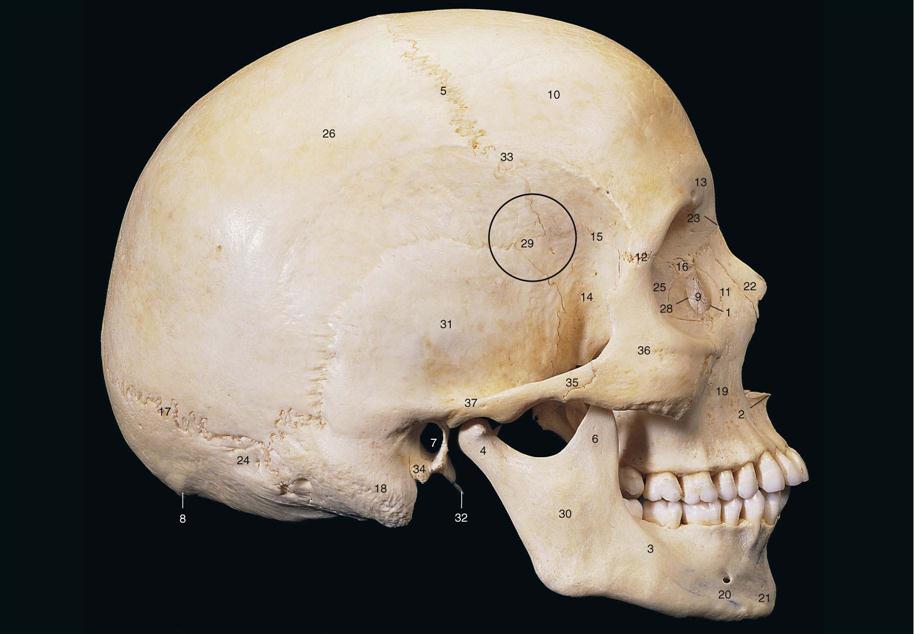
6 Coronoid process of mandible
7 External acoustic meatus of temporal bone
8 External occipital protuberance (inion)
10 Frontal bone
13 Glabella
14 Greater wing of sphenoid bone
18 Mastoid process of temporal bone
19 Maxilla
22 Nasal bone
23 Nasion
25 Orbital part of ethmoid bone
27 Pituitary fossa (sella turcica)
31 Squamous part of temporal bone
32 Styloid process of temporal bone
(From Abrahams PH, Marks SC Jr, Hutchings RT: McMinn’s color atlas of human anatomy, ed 5, St Louis, 2003, Mosby.)
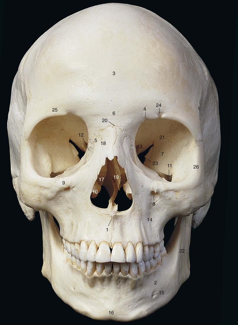
Frontal Bone
The frontal bone forms the forehead, part of the floor of the cranium, and most of the roof of the orbits. (The orbit is the bony cavity that protects the eye.) The frontal bone contains the two frontal sinuses, with one located above each eye (Fig. 9-3).
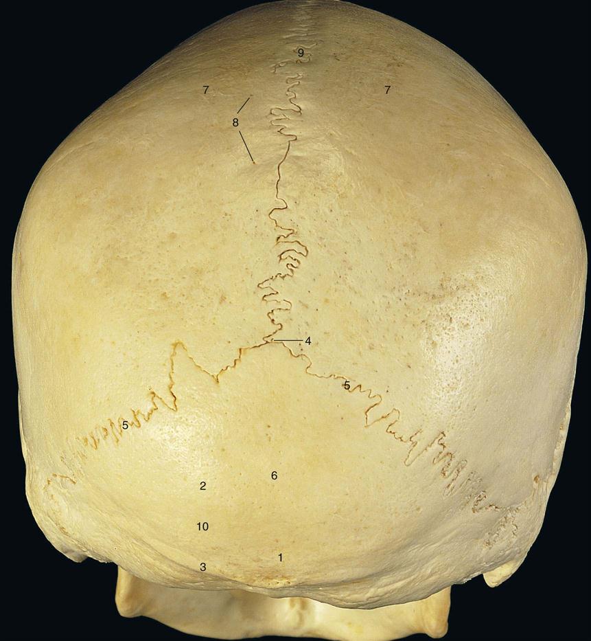
Occipital Bone
The occipital bone forms the back and base of the cranium (Fig. 9-4). It joins the parietal bones at the lambdoid suture. The spinal cord passes through the foramen magnum of the occipital bone.
Temporal Bones
Paired temporal bones form the sides and base of the cranium (see Fig. 9-2). Each temporal bone encloses an ear and contains the external auditory meatus, which is the bony passage of the outer ear.
The mastoid process is a projection on the temporal bone located just behind the ear. The mastoid process is composed of air spaces that communicate with the middle ear cavity.
The lower portion of each temporal bone bears the glenoid fossa for articulation with the mandible. The styloid process extends from the undersurface of the temporal bone.
Sphenoid Bone
The sphenoid bone is made up of a body and paired greater and lesser wings. It forms the anterior part of the base of the skull (see Fig. 9-2).
Each greater wing articulates with the temporal bone on either side and anteriorly with the frontal and zygomatic bones to form part of the orbit. Each lesser wing articulates with the ethmoid and frontal bones and also forms part of the orbit.
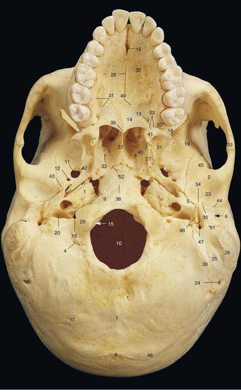
The sphenoid sinuses are located in the sphenoid bone just posterior to the eye. The pterygoid process, which extends downward from the sphenoid bone, consists of two plates. The lateral pterygoid plate is the point of origin for the internal and external pterygoid muscles. The medial pterygoid plate ends in the hook-shaped hamulus (Fig. 9-5), which is visible on some dental radiographs.
Stay updated, free dental videos. Join our Telegram channel
VIDEdental - Online dental courses


