Oral Embryology and Histology
Learning Outcomes
On completion of this chapter, the student will be able to achieve the following objectives:
• Pronounce, define, and spell the Key Terms.
• Define embryology and histology.
• Describe the three periods of prenatal development.
• Discuss prenatal influences on dental development.
• Describe the functions of osteoclasts and osteoblasts.
• Describe the steps in the formation of the palate.
• Describe the stages in the development of a tooth.
• Discuss genetic and environmental factors that can affect dental development.
• Discuss the life cycle of a tooth.
• Explain the differences between clinical and anatomic crowns.
• Name and describe the tissues of the teeth.
• Name and describe the three types of dentin.
• Describe the structure and location of dental pulp.
• Name and describe the components of the periodontium.
• Describe the functions of periodontal ligaments.
• Describe the various types of oral mucosa and give an example of each.
Electronic Resources
 Additional information related to content in Chapter 8 can be found on the companion Evolve Web site.
• Labeling Exercise: Identify the Dental Tissues of an Anterior Tooth
• Labeling Exercise: Identify the Dental Tissues of a Posterior Tooth
• WebLinks
Key Terms
Alveolar crest Highest point of the alveolar ridge.
Alveolar socket Cavity within the alveolar process that surrounds the root of a tooth.
Ameloblasts (uh-MEL-oe-blasts) Cells that form enamel.
Anatomic crown Portion of the tooth that is covered with enamel.
Apex Tapered end of each root tip.
Apical foramen Natural opening in the root.
Cementoblasts (se-MEN-toe-blasts) Cells that form cementum.
Cementoclasts (se-MEN-toe-klasts) Cells that resorb cementum.
Cementum Specialized, calcified connective tissue that covers the anatomic root of a tooth.
Clinical crown That portion of the tooth that is visible in the oral cavity.
Conception Union of the male sperm and the female ovum.
Coronal pulp Part that lies within the crown portion of the tooth.
Cortical plate Dense outer covering of spongy bone that makes up the central part of the alveolar process.
Dental lamina Thickened band of oral epithelium that follows the curve of each developing arch.
Dental papilla Gingivae between the teeth.
Dental sac Connective tissue that envelops the developing tooth.
Dentinal fiber Fibers found in dentinal tubules.
Dentinal tubules Microscopic canals found in dentin.
Deposition The process by which the body adds new bone.
Embryo An organism in the earliest stages of development.
Embryology (em-bree-OL-uh-jee) The study of prenatal development.
Embryonic (em-bree-ON-ik) period Stage of human development that occurs from the beginning of the second week to the end of the eighth week.
Enamel organ Part of a developing tooth destined to produce enamel.
Enamel spindles The ends of odontoblasts (dentin-forming cells) that extend across the detinoenamel junction a short distance into the enamel.
Exfoliation (eks-foe-lee-AY-shun) The normal process of shedding the primary teeth.
Fetal period Stage of human development that starts at the beginning of the ninth week and ends at birth.
Gestation (jes-TAY-shun) Stage of human development that starts at fertilization and ends at birth.
Histology (his-TOL-uh-jee) The study of the structure and function of body tissues on a microscopic level.
Hunter-Schreger bands Alternating light and dark bands in the enamel that are produced when enamel prisms intertwine or change direction.
Hydroxyapatite Mineral compound that is the principal inorganic component of bone and teeth.
Lamina dura Thin, compact bone that lines the alveolar socket; also known as the cribriform plate.
Mandibular (man-DIB-yoo-lur) arch The lower jaw.
Masticatory (MAS-ti-kuh-tor-ee) mucosa Oral mucosa that covers the hard palate, dorsum of the tongue, and gingiva.
Meiosis (mye-OE-sis) Reproductive cell production that ensures the correct number of chromosomes.
Odontoblasts (o-DON-to-blasts) Cells that form dentin.
Odontogenesis (o-don-to-JEN-eh-sis) Formation of new teeth.
Osteoblasts (OS-tee-oe-blasts) Cells that form bone.
Osteoclasts (OS-tee-oe-klasts) Cells that resorb bone.
Periodontium (per-ee-oe-DON-shee-um) Structures that surround, support, and are attached to the teeth.
Preimplantation period Stage of development that occurs during the first week after fertilization.
Prenatal (pree-NAY-tul) development Stage of human development that starts at pregnancy and ends at birth.
Primary cementum Cementum that covers the root of the tooth and is formed outward from the cementodentinal junction for the full length of the root.
Primary dentin Dentin that forms before eruption and that makes up the bulk of the tooth.
Primary palate The shelf that separates the oral and nasal cavities.
Prism A calcified column or rod.
Pulp chamber The space occupied by pulp.
Radicular pulp The other portion of pulp known as root pulp.
Remodeling Growth and change in shape of existing bone that involves deposition and resorption of bone.
Resorption (ree-SORP-shun) The body’s processes of eliminating existing bone or hard tissue structure.
Secondary cementum Cementum that is formed on the apical half of the root; also known as cellular cementum.
Secondary dentin Dentin that forms after eruption and continues at a very slow rate throughout the life of the tooth.
Secondary palate The final palate formed during embryonic development.
Specialized mucosa Mucous membrane on the tongue in the form of lingual papillae, which are structures associated with sensations of taste.
Stomodeum The primitive mouth.
Stratified squamous (SKWA-mus) epithelium Layers of flat, formed epithelium.
Striae of Retzius Incremental rings that represent variations in deposition of the enamel matrix during tooth formation.
Succedaneous (suk-se-DAY-ne-us) teeth Permanent teeth that replace primary teeth.
Tertiary dentin Dentin that forms in response to irritation and appears as a localized deposit on the wall of the pulp chamber; also known as reparative dentin.
Tooth buds Enlargements produced by the formation of dental lamina.
Zygote Fertilized egg.
Embryology is the study of prenatal development throughout the stages before birth. The first part of this chapter discusses development with emphasis on the formation of the teeth and structures of the oral cavity. Learning about the development of the oral structures provides the foundation for later understanding of developmental problems that may occur in these structures.
Histology is the study of the structure and function of tissues on a microscopic level. The second part of this chapter covers the histology of the teeth, their supporting structures, and the oral mucosa, which surrounds the teeth and lines the mouth. By understanding the histology of oral tissues, the dental assistant can better understand the disease processes that occur in the oral cavity.
Oral Embryology
Pregnancy begins with conception, which is also known as fertilization. Gestation, the period from fertilization to birth, has an average duration of 9 months from conception to birth, or 40 weeks from the last menstrual period (LMP). The due date is usually figured out with a convenient rule of thumb; count back three months from the day the LMP began, then add a year and a week. The date you come up with is just a guide; the baby may arrive anywhere from two weeks earlier to two weeks later. The sex of the baby is established at conception and will be apparent in a few weeks. This little 1-inch being weighs only about .04 ounce (1 gram) and could fit comfortably on a tablespoon.
A physician usually describes prenatal development in weeks on the basis of the date of the LMP. In embryology, developmental age is based on the date of conception, which is assumed to have occurred two weeks after the LMP. Developmental ages noted in this chapter are based on the date of conception.
Prenatal Development
Prenatal development consists of three distinct periods: (1) preimplantation, (2) embryonic, and (3) fetal (Fig. 8-1).
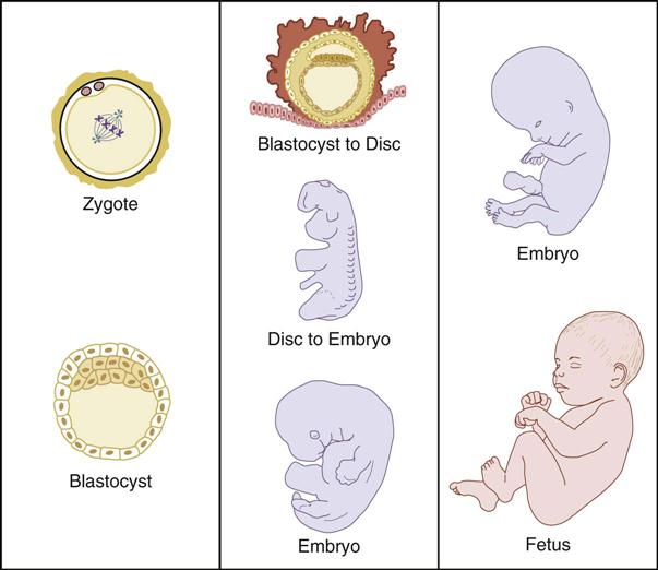
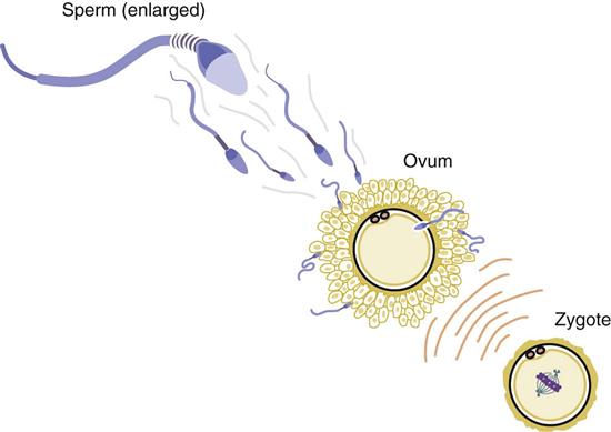
The preimplantation period takes place during the first week. At the beginning of the week, an ovum (egg) is penetrated by and united with a sperm during fertilization (Fig. 8-2). This penetration of the egg by a sperm cell has an immediate effect on the surface of the egg: The outer coating of the egg changes, so that no other sperm cell can enter. The union of the egg and the sperm subsequently forms a fertilized egg, or zygote. In the zygote, 23 chromosomes in the sperm unite with 23 chromosomes in the egg, providing a new life with a full complement of 46 chromosomes. These chromosomes will determine its inherited characteristics and will direct its growth and development. The process of joining the parents’ chromosomes is called meiosis. Meiosis ensures that the future embryo will have the correct number of chromosomes.
The embryonic period extends from the beginning of the second week to the end of the eighth week, and the new individual is known as an embryo. This period is the most critical time because during these weeks, development begins in all major structures of the body. The cells begin to proliferate (increase in number), differentiate (change into tissues and organs), and integrate (form systems). Many of these key developments occur before the mother even knows that she is pregnant. At about the end of the eighth week of pregnancy, a baby graduates from embryo to fetus. This name change signifies a change in the baby’s level of development. While an embryo, the baby looks very much like a tadpole, but as a fetus, it has a distinctly human appearance.
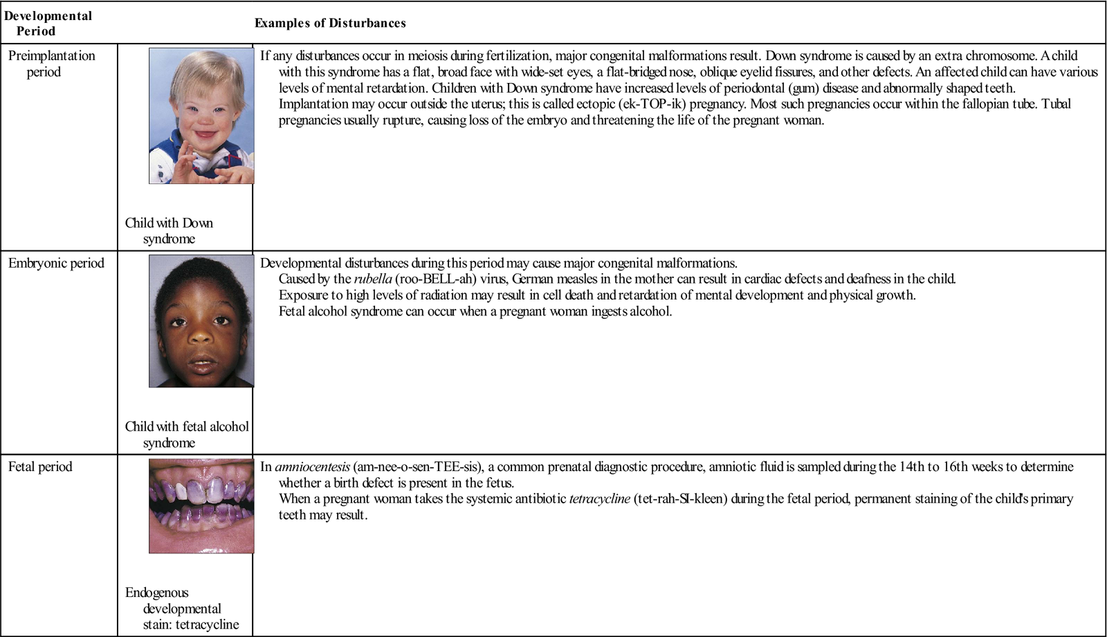
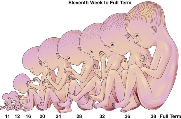
The fetal period continues from the ninth week and lasts until birth. During the fetal phase, body systems continue to develop and mature. The fetus has distinguishable ears, arms, hands, legs, and feet, as well as the fingerprints and footprints that will set it apart from other human beings. Because all organ systems are formed during the embryonic period, the fetus is less vulnerable than the embryo to malformations caused by radiation, viruses, and drugs (Table 8-1). The fetal stage is a period of growth and maturation (Fig. 8-3).

TABLE 8-1
< ?comst?>
| Developmental Period | Examples of Disturbances | |
| Preimplantation period |
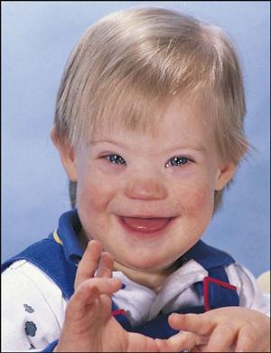 Child with Down syndrome |
If any disturbances occur in meiosis during fertilization, major congenital malformations result. Down syndrome is caused by an extra chromosome. A child with this syndrome has a flat, broad face with wide-set eyes, a flat-bridged nose, oblique eyelid fissures, and other defects. An affected child can have various levels of mental retardation. Children with Down syndrome have increased levels of periodontal (gum) disease and abnormally shaped teeth. Implantation may occur outside the uterus; this is called ectopic (ek-TOP-ik) pregnancy. Most such pregnancies occur within the fallopian tube. Tubal pregnancies usually rupture, causing loss of the embryo and threatening the life of the pregnant woman. |
| Embryonic period |
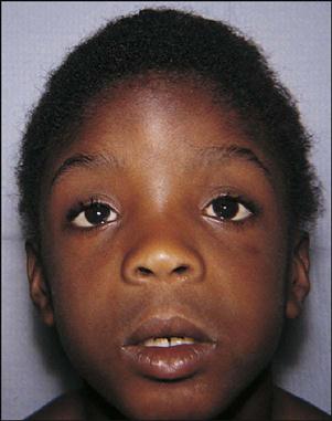 Child with fetal alcohol syndrome |
Developmental disturbances during this period may cause major congenital malformations. Caused by the rubella (roo-BELL-ah) virus, German measles in the mother can result in cardiac defects and deafness in the child. Exposure to high levels of radiation may result in cell death and retardation of mental development and physical growth. Fetal alcohol syndrome can occur when a pregnant woman ingests alcohol. |
| Fetal period |
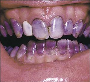 Endogenous developmental stain: tetracycline |
In amniocentesis (am-nee-o-sen-TEE-sis), a common prenatal diagnostic procedure, amniotic fluid is sampled during the 14th to 16th weeks to determine whether a birth defect is present in the fetus. When a pregnant woman takes the systemic antibiotic tetracycline (tet-rah-SI-kleen) during the fetal period, permanent staining of the child’s primary teeth may result. |
< ?comen?>< ?comst1?>
< ?comst1?>
< ?comen1?>
Figures from Zitelli BJ, Davis HW: Atlas of pediatric physical diagnosis, ed 5, St Louis, 2008, Mosby; and Daniel SJ, Harfst SA, Wilder R: Mosby’s dental hygiene: concepts, cases, and competencies, ed 2, St Louis, 2008, Mosby. (Courtesy Dr. George Taybos, Jackson, MS.)
Embryonic Development of the Face and Oral Cavity
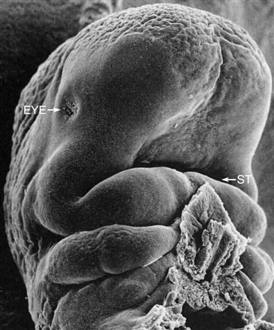
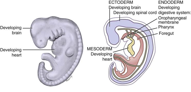
The face and its related tissues begin to form during the fourth week of prenatal development within the embryonic period. During this time, the rapidly growing brain of the embryo bulges over the oropharyngeal membrane, beating heart, and stomodeum (Fig. 8-4).
Primary Embryonic Layers
During the third week of development, the cells of the embryo form the three primary embryonic layers: ectoderm, mesoderm, and endoderm. The cells within each layer multiply and differentiate into specialized cells needed to form the organs and tissues of the body.

Early Development of the Mouth
During the fourth week, the stomodeum (primitive mouth) and the primitive pharynx merge, and the stomodeum extends into part of the mouth. By the beginning of the fifth week, the embryo is approximately 5 mm. The heart is prominent and bulging (Fig. 8-5).
The site of the face is indicated from above by the region just in front of the bulging forebrain (future forehead) and from below by the first pair of branchial arches (future jaws).
Branchial Arches
By the end of the fourth week, six pairs of branchial arches have formed. The term pharyngeal arches is often used instead of branchial arches in discussions of these tissues. The first two of these arches give rise to the structures of the head and neck (see Chapter 9).
The first branchial arch, also known as the mandibular arch, forms the bones, muscles, and nerves of the face. The first arch also forms the lower lip, the muscles of mastication, and the anterior portion of the alveolar process of the mandible.
The second branchial arch, also known as the hyoid arch, forms the styloid process, stapes of the ear, stylohyoid ligament, and part of the hyoid bone. The second arch also forms the side and front of the neck, as well as some muscles of facial expression.
The third branchial arch forms the body of the hyoid and the posterior tongue. The fourth, fifth, and sixth branchial arches form the structures of the lower throat, including the thyroid cartilage, and the muscles and nerves of the pharynx and larynx.

Stay updated, free dental videos. Join our Telegram channel
VIDEdental - Online dental courses


